Download
1 / 27
270 likes | 471 Views
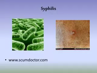
SYPHILIS. SYPHILIS INTRODUCTION. Caused by Treponema pallidum . Transmission: sexual; maternal-fetal, and rarely by other means. Primary and secondary syphilis in the US dropped by ~ 90 %t from 1990 to 2000, the number of cases have gone up since then.

E N D
SYPHILISINTRODUCTION • Caused by Treponema pallidum. • Transmission: sexual; maternal-fetal, and rarely by other means. • Primary and secondary syphilis in the US dropped by ~ 90 %t from 1990 to 2000, the number of cases have gone up since then. • A dramatic increase in cases in men from 2000 to 2002 reflected syphilis in MSM. • Syphilis increases the risk of both transmitting and getting infected with HIV. • Do HIV testing in all patients with syphilis.
STAGES OF SYPHILIS • Primary • Secondary • Latent • Early latent • Late latent • Late or tertiary • May involve any organ, but main parts are: • Neurosyphilis • Cardiovascular syphilis • Late benign (gumma)
PRIMARY SYPHILIS(The Chancre) • Incubation period 9-90 days, usually ~21 days. • Develops at site of contact/inoculation. • Classically: single, painless, clean-based, indurated ulcer, with firm, raised borders. Atypical presentations may occur. • Mostly anogenital, but may occur at any site (tongue, pharynx, lips, fingers, nipples, etc...) • Non-tender regional adenopathy • Very infectious. • May be darkfield positive but serologically negative. • Untreated, heals in several weeks, leaving a faint scar.
SECONDARY SYPHILIS • Seen 6 wks to 6 mos after primary chancre • Usually diffuse non-pruritic, indurated rash, including palms & soles. • May also cause: • Fever, malaise, headache, sore throat, myalgia, arthralgia, generalized lymphadenopathy • Hepatitis (10%) • Renal: an immune complex type of nephropathy with transient nephrotic syndrome • Iritis or an anterior uveitis • Bone: periostitis • CSF pleocytosis in 10 - 30% (but, symptomatic meningitis is seen in <1%)
SECONDARY SYPHILIS(Cont.) • The skin rash: • Diffuse, • often with a superficial scale (papulosquamous). • May leave residual pigmentation or depigmentation. • CondylomataLata: • Formed by coalescence of large, pale, flat-topped papules. • Occur in warm, moist areas such as the perineum. • Highly infectious. • Mucosal lesions: ~ 30% of secondary syphilis patients develop mucous patch (slightly raised, oval area covered by a grayish white membrane, with a pink base that does not bleed). • Highly infectious
LATENT SYPHILIS Positive syphilis serology without clinical signs of syphilis (& has normal CSF). • It begins with the end of secondary syphilis and may last for a lifetime. • Pt may or may not have a h/o primary or secondary syphilis. • Diseases known to cause occasional false-positive nontreponemal test reactions for syphilis, such as systemic lupus erythematosus (SLE), and congenital syphilis must be excluded before the diagnosis of latent syphilis can be made. • Is divided into early and late latency.
LATENT SYPHILIS (cont.) • Early latent: • The first year after the resolution of primary or secondary lesions, or • A reactive serologic test for syphilis in an asymptomatic individual who has had a negative serologic test within the preceding year. • Infectious. • Late latent: • Usually not infectious, except for the pregnant woman, who may transmit infection to her fetus.
LATE SYPHILIS‘Tertiary Syphilis’ • Is the destructive stage of the disease. • Lesions develop in skin, bone, & visceral organs (any organ). • The main types are: • Late benign (gummatous) • Cardiovascular & • Neurosyphilis • Can be crippling and life threatening • Blindness, deafness, deformity, lack of coordination, paralysis, dementia may occur • It is usually very slowly progressive, barring certain neurologic syndromes which may develop suddenly due to endarteritis and thrombosis in the CNS • Late syphilis is noninfectious.
LATE BENIGN SYPHILIS (THE GUMMA) • The gumma was the most common complication of late syphilis in the Oslo Study of untreated patients (1891 to 1951); rare in the penicillin era. • Usually develop 1-10 years after infection and may involve any part of the body. • Gummas may be single or multiple. Start as a superficial nodule or as a deeper lesion that breaks down to form punched-out ulcers. They are ordinarily indolent, slowly progressive, and indurated granulomata, with central healing with an atrophic scar surrounded by hyperpigmented borders. • Cutaneous gummas may be confused with skin lesions of TB, sarcoidosis, leprosy, and deep fungal infections (but, gumma is the only such lesion to heal dramatically with penicillin therapy). Gumma can also be papulosquamous type mimicking psoriasis. • T. pallidum is ordinarily not demonstrable by silver stain but can sometimes be recovered by inoculation of rabbits. • May be destructive, but responds rapidly to treatment, thus, is relatively benign.
TESTS FOR SYPHILIS • Dark field Microscopy • VDRL, RPR • FTA-ABS, MHA-TP • Direct Fluorescent Antibody (DFA)
Primary, Secondary, Early LatentSyphilis Recommended regimen -Benzathine Penicillin G, 2.4 million units IM Penicillin Allergy* -Doxycycline 100 mg twice daily x 14 days or -Ceftriaxone 1 gm IM/IV daily x 8-10 days (limited studies) or -Azithromycin 2 gm single oral dose (preliminary data) *
Congenital Syphilis • Congenital syphilis is transmitted in utero after the first 16 weeks of pregnancy, therefore it is usually not a cause of abortion during the first trimester. • The infected child born later in a family usually has less severe syphilis. • Again, it has been divided according to the arbitrary dividing line of two years into early and late types. • Early Congenital • The features are similar to secondary syphilis. Usually it occurs 2-8 weeks after birth, presenting with failure to thrive, muco-cutaneous lesions (condylomatalata), generalized lymphadenopathy, nasal snuffles and skin rash. • Late Congenital • The onset usually occurs at or near puberty. Well-known stigmata include nerve deafness, interstitial keratitis, Hutchison's teeth (Hutchison's triad), rhagades around mouth, clutton's joint, osteitis & chondritis (saddle nose, frontal bossing, sabre tibia) and perforated palate.
CONGENITAL SYPHILIS • The permanent dentition may show characteristic abnormalities known as Hutchinson's teeth; the upper central incisors are widely spaced, centrally notched, and tapered in the manner of a screwdriver. The molars may show multiple, poorly developed cusps (i.e., mulberry molars).
Congenital SyphilisInfants with Seroreactive Mothers • Nontreponemal test on infant serum • Examination (nonimmune hydrops, jaundice, rhinitis, rash) • Pathologic exam of placenta or umbilical cord (fluorescent antitreponemal antibody) • Darkfield or DFA of suspicious lesions or body fluids
Congenital SyphilisProven/highly probable disease Aqueous crystalline penicillin G 100,000 -150,000 units/kg/day, administered as 50,000 units/kg/dose IV q 12 hours during the first 7 days and thereafter q 8 hours for 10 days or Procaine penicillin G 50,000 units/kg/dose IM in a single daily dose for 10 days