Download
1 / 19
190 likes | 495 Views
developmental venous anomalies. veneuze angiomen Rob Rundervoort 29 juni 2005. casus 1. ♂ 55 jr. VG: hypertensie mei 2005: presentatie SEH anamnese: plotseling ontstane taalstoornis en onhandigheid rechter lichaamshelft gedurende enkele uren geen trekkingen of bewustzijnsdaling.

E N D
developmental venous anomalies veneuze angiomen Rob Rundervoort 29 juni 2005
casus 1 • ♂ 55 jr. • VG: hypertensie • mei 2005: presentatie SEH • anamnese: plotseling ontstane taalstoornis en onhandigheid rechter lichaamshelft gedurende enkele uren • geen trekkingen of bewustzijnsdaling
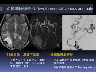
casus 1 - vervolg • neurologisch onderzoek: geen afwijkingen • conclusie: TIA linker hemisfeer • CT cerebrum:
casus 1 – vervolg • bij aanvullend MRA g.a.v. AVM • conclusie MRI: veneus angioom • lab, ECG en duplex carotiden g.a. • relatie veneus angioom met klachten?
casus 2 • ♀ 69 jaar • VG: ossaal gemetastaseerd mammaca. • hemifacialisspasmen rechts • bij neurologisch onderzoek g.a.
developmental venous anomalies • DVA = veneus angioom • meest voorkomende cerebrale vasculaire malformatie • aanlegstoornis • weinig literatuur • alle retrospectief
DVA • prevalentie: • 2.56 % (autopsie-studie) • 0.48-0.7 % (MRI-studies) • lokatie: 1/3 infratentorieel 2/3 supratentorieel
DVA en intracerebrale hematomen • bij 19% (12/67) caverneus hemangioom • alle patiënten met ICH en DVA caverneus hemangioom in bloedingsregio • DVA kan hierbij op afstand liggen
DVA en epilepsie • van 15 patiënten met epilepsie en DVA slechts één met focus in regio DVA (temporaal links) • selectieve amygdalohippocampectomie met intact laten DVA aanvalsvrij • dus: geen bewijs dat DVA geassocieerd is met epilepsie
DVA en ischemie • TIA in literatuur nooit aan DVA toegeschreven • casuïstiek: veneus infarct bij getromboseerde afvoerende vene
hypothese • veneus infarct bij patiënt 1 na doorgemaakte trombose ? • echter: geen trombus zichtbaar bij beeldvorming
conclusies • veneuze angiomen zelden van pathologische betekenis • soms geassocieerd met al dan niet symptomatisch caverneus hemangioom • in een enkel geval: veneus infarct door trombose