Download
1 / 1
10 likes | 137 Views
Human Mesenchymal Stem Cell Behavior on 3D PEDGA Superporous Hydrogels Taneka Denise Taylor, RET Fellow 2009 Morgan Park High School RET Mentor: Dr. Gemeinhart, PhD NSF- RET Program. Introduction. Abstract.

E N D
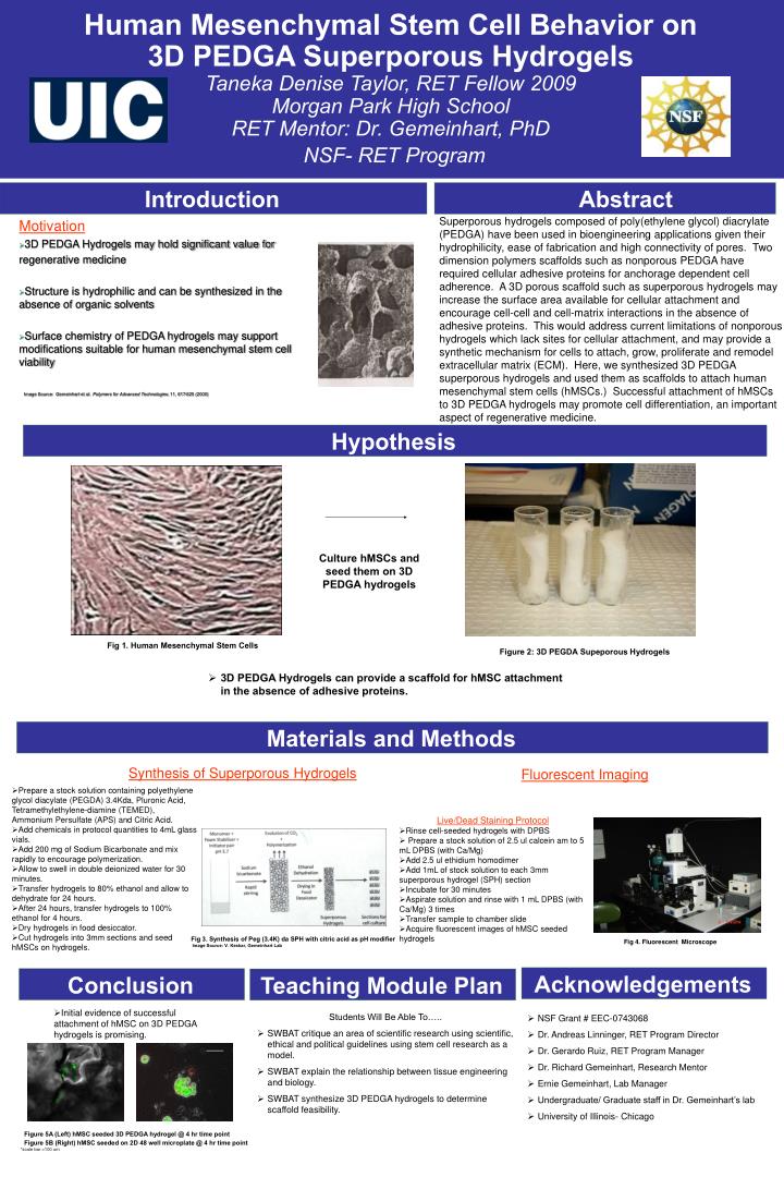
Human Mesenchymal Stem Cell Behavior on 3D PEDGA Superporous HydrogelsTaneka Denise Taylor, RET Fellow 2009Morgan Park High SchoolRET Mentor: Dr. Gemeinhart, PhDNSF- RET Program Introduction Abstract Superporous hydrogels composed of poly(ethylene glycol) diacrylate (PEDGA) have been used in bioengineering applications given their hydrophilicity, ease of fabrication and high connectivity of pores. Two dimension polymers scaffolds such as nonporous PEDGA have required cellular adhesive proteins for anchorage dependent cell adherence. A 3D porous scaffold such as superporous hydrogels may increase the surface area available for cellular attachment and encourage cell-cell and cell-matrix interactions in the absence of adhesive proteins. This would address current limitations of nonporous hydrogels which lack sites for cellular attachment, and may provide a synthetic mechanism for cells to attach, grow, proliferate and remodel extracellular matrix (ECM). Here, we synthesized 3D PEDGA superporous hydrogels and used them as scaffolds to attach human mesenchymal stem cells (hMSCs.) Successful attachment of hMSCs to 3D PEDGA hydrogels may promote cell differentiation, an important aspect of regenerative medicine. • Motivation • 3D PEDGA Hydrogels may hold significant value for • regenerative medicine • Structure is hydrophilic and can be synthesized in the absence of organic solvents • Surface chemistry of PEDGA hydrogels may support modifications suitable for human mesenchymal stem cell viability • Image Source: Gemeinhart et al. Polymers for Advanced Technologies. 11, 617-625 (2000) Hypothesis Hypothesis Culture hMSCs and seed them on 3D PEDGA hydrogels Fig 1. Human Mesenchymal Stem Cells Figure 2: 3D PEGDA Supeporous Hydrogels • 3D PEDGA Hydrogels can provide a scaffold for hMSC attachment in the absence of adhesive proteins. Materials and Methods Synthesis of Superporous Hydrogels Fluorescent Imaging • Prepare a stock solution containing polyethylene glycol diacylate (PEGDA) 3.4Kda, Pluronic Acid, Tetramethylethylene-diamine (TEMED), Ammonium Persulfate (APS) and Citric Acid. • Add chemicals in protocol quantities to 4mL glass vials. • Add 200 mg of Sodium Bicarbonate and mix rapidly to encourage polymerization. • Allow to swell in double deionized water for 30 minutes. • Transfer hydrogels to 80% ethanol and allow to dehydrate for 24 hours. • After 24 hours, transfer hydrogels to 100% ethanol for 4 hours. • Dry hydrogels in food desiccator. • Cut hydrogels into 3mm sections and seed hMSCs on hydrogels. • Live/Dead Staining Protocol • Rinse cell-seeded hydrogels with DPBS • Prepare a stock solution of 2.5 ul calcein am to 5 mL DPBS (with Ca/Mg) • Add 2.5 ul ethidium homodimer • Add 1mL of stock solution to each 3mm superporous hydrogel (SPH) section • Incubate for 30 minutes • Aspirate solution and rinse with 1 mL DPBS (with Ca/Mg) 3 times • Transfer sample to chamber slide • Acquire fluorescent images of hMSC seeded hydrogels Fig 3. Synthesis of Peg (3.4K) da SPH with citric acid as pH modifier Image Source: V. Keskar, Gemeinhart Lab Fig 4. Fluorescent Microscope Acknowledgements Conclusion Teaching Module Plan • Initial evidence of successful attachment of hMSC on 3D PEDGA hydrogels is promising. • Students Will Be Able To….. • SWBAT critique an area of scientific research using scientific, ethical and political guidelines using stem cell research as a model. • SWBAT explain the relationship between tissue engineering and biology. • SWBAT synthesize 3D PEDGA hydrogels to determine scaffold feasibility. • NSF Grant # EEC-0743068 • Dr. Andreas Linninger, RET Program Director • Dr. Gerardo Ruiz, RET Program Manager • Dr. Richard Gemeinhart, Research Mentor • Ernie Gemeinhart, Lab Manager • Undergraduate/ Graduate staff in Dr. Gemeinhart’s lab • University of Illinois- Chicago Figure 5A (Left) hMSC seeded 3D PEDGA hydrogel @ 4 hr time point Figure 5B (Right) hMSC seeded on 2D 48 well microplate @ 4 hr time point *scale bar =100 um