Download
1 / 41
500 likes | 1.54k Views
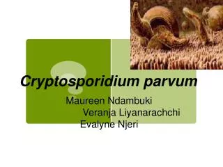
Toxoplasma gondii & Cryptosporidium parvum. Both belong to phylum Apicomplexa. Both are sporozoites. Infection is very widespread. Morphology. Important stages in life cycle - Tachyzoites (intra cellular trophozoites or proliferative forms) Tissue cysts Oocysts

E N D
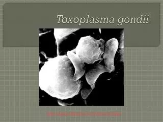
Toxoplasma gondii & Cryptosporidium parvum Both belong to phylum Apicomplexa. Both are sporozoites. Infection is very widespread.
Morphology Important stages in life cycle - • Tachyzoites (intra cellular trophozoites or proliferative forms) • Tissue cysts • Oocysts All are infectious to man.
Crescent shaped with pointed anterior end & rounded posterior end. 6 μ x 2 μ Groups of proliferating tachyzoites within host cells are called Pseudocyst
Tissue cyst • Occur in chronic inf • Formed when parasite multiplies and produces wall within host cell • Bradyzoites are slowly multiplying forms seen in tissue cysts • Size – 5 μ to 100 μ
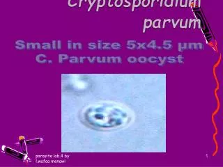
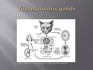
Oocyst • Seen in cats and other felines. Not seen in man • Oval or spherical 10-12 μ, contains sporoblast • Wall has two colourless layers
Life cycle • Definitive host – Cat & felines • All the forms occur viz Tachyzoites, tissue cysts, Oocyst ie schizogony & gametogony occur • Intermediate host – Other mammals incl Man and birds – Only asexual forms occur viz Tachyzoites, tissue cysts • Tachyzoites & tissue cysts represent asexual multiplication (Schizogony) • Oocyst is formed by sexual reproduction (gametogony)
Specimens – Blood (buffy coat of heparinised blood), sputum, BM, CSF, exudates, LN, spleen, Brain biopsy, vesicular fluid. Microscopic Exam – Smears/sections stained with Giemsa/PAS show parasite. Animal inoculation – IP inoculation in mice. Usually death in 4-6 weeks. Confirm by tissue cysts in brain. Lab diagnosis
Inoculation of tissue culture PCR detection of toxoplasmal DNA Serology – Detection of Ab. Sabin-Feldman dye test uses lab cultured toxoplasma. IHA Indirect fluorescent assays Latex agglutination tests ELISA Paired samples showing 16 fold rise in Ab titre indicates acute infection. Lab diagnosis (contd…)
Prevention • Avoidance of human contact specially pregnant women with cat faeces / uncooked meat • Proper cooking of meat • Hand washing, proper cleaning of chopping boards, knives etc.
Mode of inf – Direct contact with infected animal or contaminated food or water. Oocyst not destroyed by chlorination Produces mild self limited diarrhoea. In immuno compromised can cause cholera like diarrhoea of severe nature. 3-6 or even 17 litres of water lost per day. Seen in 2-5% of AIDS cases Pathogenecity
Sample – Stool Wet mount highly refractile spherical oocysts Concentration technique – ZnSO4 / Formal ether Staining – Acid fast stain, IFA Serology – IFA, ELISA Lab diagnosis
Coccidia and Microsporidia – Cause diarrhea and when they infect people with an immune suppressed state. Mature trophozoites (the feeding phase) and sexual and asexual reproduction occur within epithelial cells in a single host. – Called -“one stop shopping.” Oocysts are then excreted into the environment. The oocysts can immediately or, if a period of maturation is needed, eventually infect another host.
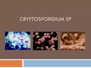
Coccidians Important in HIV/AIDS • Cryptosporidium parvum and Cryptosporidium hominis • Cyclospora cayetanensis • Isospora belli • Microsporidia • Obligate intracellular spore forming protozoa consisting of hundreds of species and causing symptoms almost exclusively in AIDS patients.
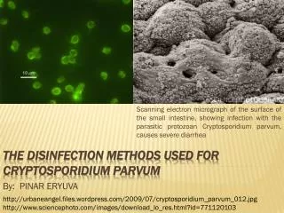
Cryptosporidium • parvum and hominis • Species: Many • Inoculum – Median • 132 oocysts. • 3) Source of parasite – • Person to person. Water – both drinking • and recreational. • Animal or human • feces contaminates • water sources. Oocysts infective when passed. Infected while swimming CDC
In 1993 waterborne cryptosporidiosis occurred in Milwaukee. Cryptosporidium is highly resistant to chlorine and difficult to filter out. Spring rains washed animal waste into drinking water sources. More than 400,000 residents were affected; 103 people died. Milwaukee NASA
Cyclospora cayetanensis • Species: Human • (All human cyclosporiasis is caused by Cyclospora cayetanensis.) • 2) Inoculum: – ? • 3) Source: Basil, • Raspberries, Water
Invasiveness: Small bowel epithelial cells • where replication takes place. • Incubation period: 1 week • Clinical manifestations: Variable: Asymptomatic to secretory diarrhea with dehydration, abdominal pain, nausea, anorexia, fever, and weight loss. • Usually self-limited subsiding within 2 weeks if CD4+ count > 180. Prolonged (years) 25 L/d watery diarrhea if CD4+ count < 150. • Cholecystitis in the HIV/AIDS patient.
Sporulation at day 5: Since the cysts are not sporulated when passed in the feces, direct person to person spread does not occur. CDC 4) Invasion: Small bowel epithelial cells 5) Incubation: – 1 week
Cyclospora cayetanensis 6) Clinical Manifestations:Secretory diarrhea without blood or WBC; anorexia, nausea, weight loss. Relapsing or persisting for 3 months. Cholecystitis in HIV/AIDS. 7) Laboratory: Stool microscopy. Not found on “Stool for O&P” (Needs modified acid fast.) Cyclospora Cryptosporidium
8) Treatment: TMP-SMX, Quinolone 9) Prophylaxis: Peel, wash; It can come here. 10) Travelers: Peel, wash; You can get it there. CDC Basil USDA Raspberries contaminated with Cyclospora.
Cryptosporidium, Cyclospora, and Isospora all cause a secretory diarrhea without RBC or WBC in the stool. None are easily found in routine “stool for ova and parasites.” Modified acid-fast staining (no heating of reagents needed) is useful for all of the Coccidians. Isospora belli Cyclospora Cryptosporidium CDC
Other sporozoa • Babesia – Intra erythrocytic parasite seen in sheep & cattle. Transmitted to man by tick bite. • Pneumocystis carinii – Lives in lung alveoli. Demonstration – Silver methenamine stain.