Download
1 / 37
690 likes | 1.56k Views
Ocular Diseases of Companion Animals. Animal Technology VT-116 Adapted from Dr. Kristek. The Eyelids. Serve to protect, clean, and keep the globe hydrated Common diseases: Entropion Hair chronically touches cornea

E N D
Ocular Diseases of Companion Animals Animal Technology VT-116 Adapted from Dr. Kristek
The Eyelids • Serve to protect, clean, and keep the globe hydrated • Common diseases: • Entropion • Hair chronically touches cornea • Shar Pei, Chow, Bulldog, Golden Retriever, Persian cats, also seen in lambs and kids • Can be hereditary or acquired: spastic entropion secondary to pain • Ectropion • Exposure of conjunctiva leads to inflammation and higher risk of infection • St. Bernards, spaniels, hounds
The Eyelids • Entropion • Ectropion
The Eyelids • Meibomian Glands – aka Tarsal Glands • Produce meibum, a lipid-heavy secretion that slows down tear evaporation and increases surface tension, holding tears in-between eyelids and surface of eyeball
The Eyelids • Meibomian Gland Adenoma • Most common form of neoplasia in the eyelid in dogs • Nearly always benign • Most common in dogs > 8 years old • Other eyelid tumors • Melanoma, basal cell carcinoma, squamous cell carcinoma (SCC), adenomas, papillomas
Conjunctiva • Conjunctivitis – inflammation of the conjunctiva • Most common eye disease in domestic animals • Can be due to infection, trauma, or allergic response • Common signs: • Erythema • Discharge • Inflammation • Discomfort (demonstrated by repeated blinking, squinting, or abnormal head position)
The Cornea • Corneal ulcer – a break or erosion in the layers that form the cornea
The Cornea • Corneal ulcer • Can be the result of trauma, infection, or of idiopathic causes • Can affect one or more of the five layers of the cornea: • Epithelium • Bowman’s Layer/Bowman’s Membrane • Stroma • Descemet’s membrane • Endothelium
The Cornea • Corneal ulcer • Not usually visible to the naked eye • Fluorescein eye stain test • Two categories: • Simple ulcers • Deep Ulcers • http://www.youtube.com/watch?v=3dr1FQB6Va8
The Cornea • Keratoplasty • A graft of conjunctiva or flap of donor corneal tissue is placed over the site of the descemetocele. • The graft provides immediate blood supply to damaged or abnormal cornea to speed healing and increase the amount of medications reaching the area
The Cornea • Corneal Debridement • Debridement of the ulcer promotes healing by removing the outer non-adhering layer of the cornea with a cotton swab.
The Cornea • Grid Keratectomy • The tissue under the diseased outer surface of the cornea is altered by making tiny scratches on the surface with a fine gauge hypodermic needle.
The Cornea • Superficial Keratectomy • The outer or superficial layer of the affected cornea is surgically removed by careful dissection, using an operating microscope.
The Cornea • Pannus
Fluorescein Dye Test PROCEDURE: • Take out the Fluorescein dye strip, being careful to handle the non-dye end • Apply a drop of eye wash or saline to the orange end of the strip, and touch the strip to the animal’s eye • If only one eye appears infected, touch the normal eye first, then the infected eye • Allow the animal to blink
Fluorescein Dye Test PROCEDURE cont’d: • Rinse the eyes will with eye wash • Use a wood’s lamp, an ophthalmoscope with blue light, or even a simple pen light to examine the surface of the cornea for any retention of stain. It may be very subtle, or very obvious – so take your time examining • May wish to darken room to better visualize the stain
The Uvea • Uveitis • MANY causes, may be multifactorial • Infection • Immune-mediated reaction • Metabolic • Parasitic • Traumatic • Neoplastic
Schirmer Tear Test (STT) • Requires the use of a special tear test strip which looks like a small piece of filter paper, sometimes with a scale in millimeters printed on it • The end of the strip is bent and then carefully placed under patient’s lower eyelid • Leave the strip in this position for 60 seconds • As the tear film travels up the strip, it creates a line that can be measured on the mm scale
Schirmer Tear Test • Normal tear production in the dog is 14-24 mm/minute • A KCS diagnosis will be made with less than 10mm/min results AND clinical signs of KCS • One low measurement of tear production without clinical signs is not diagnostic of KCS
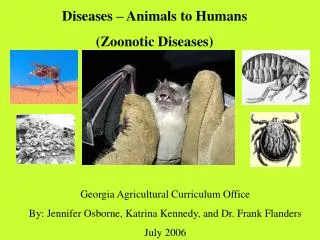
Infectious Bovine Keratoconjunctivitis “Pink Eye” in cattle • Highly Infectious, highly contagious • Pathogen is the bacteria Moraxellabovis • UV exposure and dust irritants can lead to infection • Transmitted by face flies • Tx: antibiotics, fly control, reduce dust • Nictitating membrane or eyelids may be temporarily sewn over the eye in cases of severe corneal ulceration
The Lens • Classification of cataracts: • Congenital – present at birth • Juvenile – occurs in young dogs. Usually hereditary • Diabetic – related to abnormal glucose metabolism • Senile – due to aging • Secondary to PRA, diabetes, or uveitis • Traumatic – secondary to injury • http://www.youtube.com/watch?v=yypMG4kB8zc
The Lens • Luxated Lens • The suspensory ligaments that attach lens to the ciliary break free
The Retina and Optic Nerve • Retinal Hemorrage
Open-Angle Glaucoma • In open-angle glaucoma, the drainage canals themselves are blocked, but the space between the iris and the canals are open
Closed-Angle Glaucoma • In closed-angle glaucoma, the opening to the drainage canals are blocked by another structure, the angle between iris and cornea is closed
Tonometry • Procedure: • Apply topical anesthetic to eye, one drop per eye, repeat 2 to 3 times in 3-5 minute intervals • Calibrate your tonometer • Elevate patient’s head vertically • Rest the base of the tonometer on the center of the eye and let it bear the full weight on the cornea • Take a reading off the scale, then remove the tonometer • Take 3 to 5 readings from an eye, and average your readings
Tonometry • For the Schiotz tonometer, the relationship between the IOP and the reading is inverse: the lower the reading, the higher the pressure • Normal is 3.5 to 6.5 (using the standard 5.5g weight) • If the reading is below 3.5, then additional weight is needed • Actual (converted) IOP normals: • Dog: 12.2 – 25 mmHg • Cat: 10 – 25 mmHg