Download
1 / 22
230 likes | 462 Views
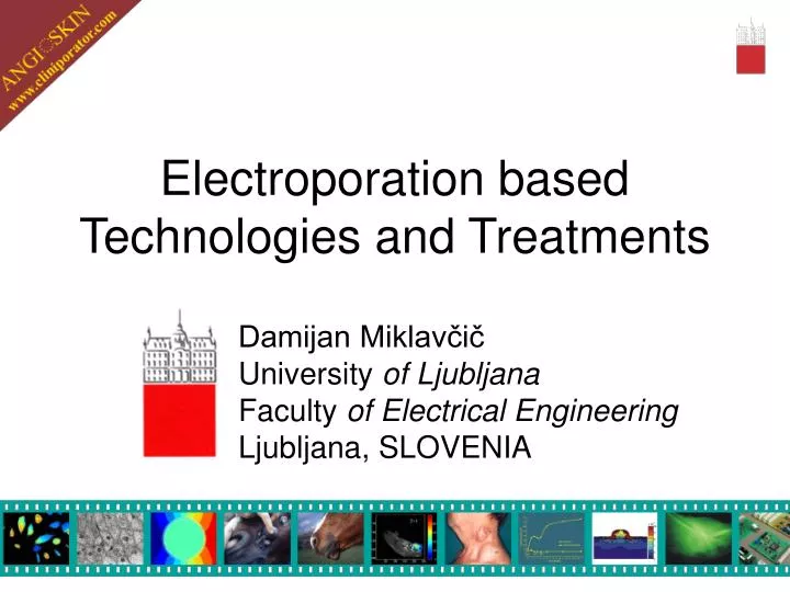
Electroporation based Technologies and Treatments. Damijan Miklavčič University of Ljubljana Faculty of Electrical Engineering Ljubljana, SLOVENIA.

E N D
Electroporation based Technologies and Treatments Damijan Miklavčič University of Ljubljana Faculty of Electrical Engineering Ljubljana, SLOVENIA
“Cells can be funny. Try to grow them with a slightly wrong recipe, and they turn over and die. But hit them with an electric field strong enough to knock over a horse, and they do enough things to justify international meetings, to fill a sizeable book, and to lead one to speak of an entirely new technology for cell manipulation.” Adrian Parsegian fromthe foreword to the Electroporation and Electrofusion in Cell Biology Edited by E. Neumann, A. S. Sowers and C. A. Jordan
Electroporation based Technologies and Treatments • Molecular cell biology research • Protein insertion into cell membrane • Cell fusion • Gene expression silencing by siRNA • Electrochemotherapy • Genetherapy based on electro genetransfer • Transdermal drug delivery • Tissue ablation • Biotechnology • Water and liquid food sterilisation
Electrochemotherapy = = chemotherapy + electroporation Electric pulse generator Chemotherapeutic surrounds the cells Increased membrane permeability allows access to the cytosol Pores reseal Electrodes time Tumour Electric pulse application Injecting chemotherapeutic
Skin metastasis of squamous cell carcinoma of the supraglottis
Skin metastasis of malignant melanoma Before treatment One year after ECT
EU 5th FP: Quality of Life Cliniporator QLK3-1999-00484 (2000-2003) • Electro-medical device for ECT ESOPE QLK3- 2002-02003 (2003-2004) • European Standard Operating Procedures for ECT
Objective Response Rate: 85% No Response: 15% 171 NODULES
GeneTherapy of diseases with altered angiogenesis: Psoriasis and Malignant Melanoma
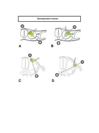
Electroporation of cells: Experiments and modelling for better understanding
Cell (electro)fusion • Monoclonal antibody production • Cancer immunotherapy • Receptor insertion into membrane • Tissue regeneration • Research (dissease models)
V homogenem električnem polju ni dielektroforeze Pozitivna dielektroforeza Negativna dielektroforeza Dielectrophoresis
Biochip and results of experiments • CHO cellls • Medium conducivity 15mS/cm • negativve dielectrofporesis • frequency 500 Hz • amplitude 4V
Electroporation based Technologies and Treatments • Molecular cell biology research • Electrochemotherapy • Electrogenetherapy • Gene expression silencing by siRNA • Transdermal drug delivery • Protein insertion into cell membrane • Cell fusion • Biotechnology • Water and liquid food sterilisation • Tissue ablation
LBK expertise and resources: Cell biology and chemistry lab Lipid bilayer setup for preparation of planar lipid membranes. • chamber for lipid bilayers • microscope with cold-light illumination for lipid bilayer observation • (SZF 30, Olympus, Germany)
LBK expertise and resources: Cell biology lab Prototype of an electroporator developed in LBK connected to digital oscilloscope LeCroy 9310C. Inverted phase contrast microscope Olympus CK40 for routine work with cell cultures & lipid vesicles.
LBK expertise and resources: Cell biology lab System for dynamic imaging of cells and lipid vesicles. • Zeiss Axiovert 200 epiflurescence • inverted microscope (Zeiss, Germany) • high resolution cooledCCD camera • (VisiCam1280) • sensitive high-speed cooled CCD • camera (BOOST 2000, Diagnostic • Instruments, GB)
List of other equipment • Other equipment available: • Spectrofluorometer (Jasco, Japan) • Microplate reader (Tecan, Switzerland) • Field jump system for fast monitoring optical changes in lipid vesicles • Computer facilities • Software: Mathematica, Matlab with different toolboxes; • Numerical modeling software: MSC/EMAS, Maxwell, COMSOL Multiphysics • NIRO2-X2: near infrared spectrophotometer for measurement of oxygenation and blood perfusion changes in tissue (Keele University, U.K.) • OxyLite 2000: 2-channel instrument for luminescence-based fiber-optic oximetry in tissue; OxyFlo 2000: 2-channel instrument for laser Doppler flowmetry in tissue; OxyData 2000: data acquisition unit for OxyLite and OxyFlo instruments (Oxford Optronix, Oxford, U.K.) • Equipment available and used in other institutions: • flow cytometer (FACSort Becton Dickinson, Mountain View, CA) Medical faculty, University ofLjubljana • Confocal microscope (Leica TCS SP5, Germany)
Collaborating Institutions SLOVENIAN: • Institute of Oncology, Ljubljana • Institute of Rehabilitation, Ljubljana • Medical Faculty, University of Ljubljana • Institute Josef Stefan, Ljubljana INTERNATIONAL: • Institut Gustave-Roussy (Villejuif, France) • Igea S.r.l. (Modena, Italy) • Universität Bielefeld (Bielefeld, Germany) • Faculty of Electrical and Computer Engineering (Zagreb, Croatia) • Institut de Pharmacologie et de Biologie Structurale (Toulouse, France) • Université catholique de Louvain (Brussels, Belgium) • Orthologic Corp. (Tempe, Arizona, USA) • Sewanee University of the South (Sewanee, Tennessee, USA) • Johns Hopkins University (Baltimore, Maryland, USA)
Laboratory for biocybernetics http://lbk.fe.uni-lj.si Peter, Katja, Marko, Jakob, Tadej, David, Matej K., Tomaž, Matej R., Denis, Blaž, Damijan, Ivan, Janez, Mojca, Stanislav, Gorazd, Vilko, Selma, Čarli, Nataša, Maša, Alenka, Barbara (mising from the photo: Anže, JernejaTina, Duša)
Thank you ! damijan.miklavcic@fe.uni-lj.si http://lbk.fe.uni-lj.si