Download
1 / 7
70 likes | 345 Views
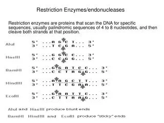
Restriction Enzymes/endonucleases. Restriction enzymes are proteins that scan the DNA for specific sequences, usually palindromic sequences of 4 to 8 nucleotides, and then cleave both strands at that position. Restriction enzymes are molecular scissors – they cut DNA

E N D
Restriction Enzymes/endonucleases Restriction enzymes are proteins that scan the DNA for specific sequences, usually palindromic sequences of 4 to 8 nucleotides, and then cleave both strands at that position.
Restriction enzymes are molecular scissors – they cut DNA • restriction enzymes are highly specific. They cut DNA only within very precise recognition sequences.
There are many different restriction enzymes that can be used. • To detect variation in DNA sequence restriction enzyme digestion can be used. • Variation in the DNA sequence that gives rise to the creation or destruction of a restriction enzyme digestion site is called a restriction fragment length polymorphism (RFLP). • Humans have two copies of each gene, except for those genes on the X and Y chromosomes in males. This must be kept in mind when doing RFLP analysis. In each sample there may be two alleles.
The original procedure used to obtain a DNA fingerprint • Isolate genomic DNA • cut with restriction enzymes • run on a gel • As humans have more than 3 billion base pairs in their genome , after electrophoresis all that can be seen is a smear because all the resulting bands overlap. • To visualise a fingerprint pattern the DNA fragments must be detected by probing and hybridisation. 4. After electrophoresis the DNA in the gel is denatured and transferred to a membrane to make a permanent record. 5. The membrane is then “probed” using a piece of sequence that is complimentary to a hypervariable region. 6. The binding of the probe is visualised using radioactivity, fluorescence, conjugated enzyme. 7. The resulting band patterns are a fingerprint. 8. The final DNA fingerprint is built by using several probes (5-10 or more) simultaneously.
Today because we have the human DNA sequence and certain other genome sequences instead of digesting total genomic DNA and creating a permanent record on a membrane that is then probed for variable regions , several different highly variable regions are amplified directly by PCR • FBI uses 22 different regions, RCMP 15 different regions, paternity tests typically use at least 7 different regions