Download
1 / 27
270 likes | 365 Views
Explore the intricate workings of circulatory systems, including portal circulation, foetal adaptations, placental functions, and the pathophysiology of shock. Learn about unique circulatory adaptations in pregnancy and foetal development.

E N D
Circulatory System (part 3) A&P 1 Tutor: Eleshia Howell
Portal circulation • Instead of the venous blood returning directly, through one capillary bed, the portal system offers a second filtering system for the blood from digestive organs. • Venous blood passes through first line capillary beds of digestive system, pancreas and spleen to go to the liver. • It then passes through secondary capillary bed in the liver (hepatic sinusoids) before entering general circulation via inferior vena cava.
The high concentration of nutrients absorbed from the stomach and intestines is first received by liver, which then regulates blood nutrient level and performs numerous chemical modifications. • The portal vein is formed by union of several other veins from organs such as spleen, intestines and stomach. It enters into inferior portion of liver. • Hepatic vein exits from posterior superior aspect of liver and empties directly into the inferior vena cava.
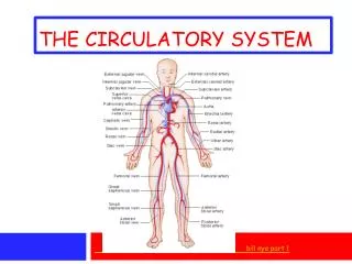
Lower body circulation P 104
Foetal circulation • The developing foetus obtains its oxygen and nutrients, and excretes its wastes, via the mother’s circulation. • Both maternal and foetal circulations develop specific adaptations unique to pregnancy. • Because the lungs, gastrointestinal system and kidneys do not begin to function until after birth, certain modifications in the foetal circulation divert flow to meet pre-natal requirements.
PLACENTA • This temporary structure provides an interface between mother and baby, allowing exchange of substances between their circulatory systems. • It develops from the surface of the fertilized ovum embedded into uterine endometrium and is expelled during the final stage of labour when it is no longer required. • It weighs about 500g, is approx 20cm diameter and about 2.5cm thick. It is firmly attached to the uterine wall and consists of an extensive network of capillaries bathed in maternal blood.
Placenta is attached to the baby via umbilical cord which contains two arteries and one vein. The cord enters the foetus at the abdomen (umbilicus). • The maternal and foetal blood supplies, while in very close proximity, are two separate circulatory systems. • Deoxygenated blood flows into the placenta from baby and is filtered by the capillary network within, removing C02 and other wastes, and returning oxygen and nutrients back into foetal circulation. • The placenta can also provide a mild protective barrier against harmful substances and some pathogens
Placenta also has essential endocrine function in that it secretes the hormones that maintain pregnancy ~ hCG, Progesterone and Oestrogen. FOETAL ADAPTATIONS: • Ductusvenosus – an extension of the umbilical vein which transports blood directly into foetal inferior vena cava, bypassing the liver. • Ductusarteriosus – small vessel connecting the pulmonary artery to the descending aorta, diverting more blood into systemic circulation (so very little blood passes through baby’s lungs).
Foramen ovale – a valve-like opening that allows blood to flow between the right and left atria, again so that most blood bypasses foetal lungs. CHANGES AT BIRTH: • When baby takes first breath and lungs inflate, pulmonary blood flow is increased. Blood returning from lungs increases pressure on left atria, closing the flap over the foramen ovale and preventing cross flow. • The increased blood O2 levels cause constriction and closure of the ductusarteriosus.
Once the placental circulation ceases soon after birth, the umbilical vein & arteries, plus ductusvenosus automatically collapse.
Pathophysiology SHOCK (circulatory failure) – occurs when the metabolic needs of cells are not being met because of inadequate blood flow. Essentially, a reduction in circulating blood volume, in blood pressure and in cardiac output. This causes tissue hypoxia, and inadequate supply of nutrients and the accumulation of waste products. There are various kinds of shock:
Hypovolaemic shock – occurs when blood volume is reduced by 15-25%. This may be caused by severe haemorrhage, extensive burns, severe vomiting or diarrhoea. • Cardiogenic shock – when damaged heart muscle cannot maintain an adequate cardiac output, eg myocardial infarction. • Septic shock – caused by severe infections in which bacterial toxins are released into the circulation, triggering a massive inflammatory and immune response. • Neurogenic shock – when a nervous system assault interferes with normal nerve control of blood vessel diameter (pain, injury, anaesthesia, severe emotional distress).
Anaphylactic shock – a severe allergic response. In the short term, there are physiological attempts to restore an adequate blood supply, eg sympathetic activation, release of ADH and renin-angiotensin-aldosterone reflex. Medical interventions may also help to restore perfusion to the brain and heart and therefore restore physiological balance. In severe cases, the cycle of hypoxia, progressive acidosis and tissue death results in organ failure, circulatory collapse, brain stem damage and ultimately death.
Embolus – a mass of any material carried in the blood, eg blood clot, tumour fragments, arterial plaque, air, pus, fat (from bone fractures), nitrogen bubbles (the ‘bends’). An embolus may interrupt blood supply or completely obstruct a vessel. • Thrombus – an intravascular blood clot which can be caused by any condition that slows blood flow, damages the smooth inner lining of the vessels or increases coagulability of blood. • Infarction – tissue death because of interrupted blood supply. • Ischaemia – tissue damage because of reduced blood supply.
Atherosclerosis – accumulation of fatty plaques in the arteries; may cause partial or complete obstruction of an artery. • Arteriosclerosis – progressive degeneration of arterial walls, associated with aging and accompanied by hypertension. • Aneurysms – abnormal local dilations of arteries; can be caused by atheroma, hypertension, defective collagen in arterial wall. If aneurysm ruptures, haemorrhage follows. • Venous thrombosis / phlebitis – inflamed thrombus in superficial or deep veins.
Varicose veins – when a vein is so dilated that the valves can not close to prevent backflow, causing a loss of elasticity and giving the appearance of elongated, tortuous and fibrous veins. This condition may be hereditary or caused by age, pregnancy, pressure, obesity, poor venous return. Most commonly occurs in the legs, but can also happen around rectum/anus (haemorrhoids), vagina / scrotum, oesophagus.
Heart failure – when the cardiac output is unable to maintain the circulation of sufficient blood to meet the needs of the body. May occur on either side of the heart (more common in left ventricle due to workload) but will eventually effect the whole organ. Over time, the heart chambers will become thicker and enlarge and the R-A-A system becomes activated leading to salt and water retention, which in turn increases blood volume and cardiac workload! • Valve disorders – heart valves may be damaged (murmurs), blocked (stenosis) or incompetent (causing regurgitation of blood).
Arrhythmias – alterations to the normal heart rate generated by the conducting system of the heart. • Bradycardia – below 60bpm • Tachycardia – above 100bpm • Asystole – no electrical activity in the ventricles, no cardiac output (flat line) • Fibrillation – disorderly contraction of the cardiac muscle fibres. AF may still produce some ineffective pumping of blood but will be rapid, irregular. VF is a medical emergency – no blood pumped into pulmonary or systemic system.
Hypertension – high blood pressure. May be of gradual onset, due to aging, linked to hereditary, lifestyle, diet, disease factors. Commonly effects the kidneys, brain, heart, blood vessels and eyes. 140/90 and beyond is considered hypertensive. • Hypotension – usually occurs as a complication to another condition. Can lead to inadequate blood supply brain. Postural hypotension is an abrupt fall in pressure on standing from a sitting / lying position which can cause dizziness / fainting (syncope).
Congenital abnormalities – present from birth. • Patent ductusarteriosus – prenatal structure fails to close completely, allowing regurgitation of blood from aorta to pulmonary artery ~ less blood in system, more in lungs ~ pulmonary congestion, heart failure. • Atrialseptal defect – “hole in the heart”; foramen ovale does not completely close off. • Fallot’sTetralogy – combination of 4 cardiac vessel / ventricle defects; surgical repair offers good prognosis.