Download
1 / 45
470 likes | 852 Views
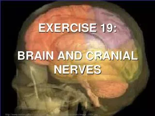
Exercise 19. The Brain & Cranial Nerves. Ventricles of the Brain. Figure 12.5. Fiber Tracts in White Matter. Figure 12.10a. Basal Nuclei. Figure 12.11a. Basal Nuclei. Figure 12.11b. Meninges.

E N D
Exercise 19 The Brain & Cranial Nerves
Ventricles of the Brain Figure 12.5
Fiber Tracts in White Matter Figure 12.10a
Basal Nuclei Figure 12.11a
Basal Nuclei Figure 12.11b
Meninges • Three connective tissue membranes lie external to the CNS – dura mater, arachnoid mater, and pia mater • Functions of the meninges • Cover and protect the CNS • Protect blood vessels and enclose venous sinuses • Contain cerebrospinal fluid (CSF) • Form partitions within the skull
Meninges Figure 12.23a
Dura Mater • Leathery, strong meninx composed of two fibrous connective tissue layers • The two layers separate in certain areas and form dural sinuses
Dura Mater • Three dural septa extend inward and limit excessive movement of the brain • Falx cerebri – fold that dips into the longitudinal fissure • Falx cerebelli – runs along the vermis of the cerebellum • Tentorium cerebelli – horizontal dural fold extends into the transverse fissure
Dura Mater Figure 12.24
Arachnoid Mater • The middle meninx, which forms a loose brain covering • It is separated from the dura mater by the subdural space • Beneath the arachnoid is a wide subarachnoid space filled with CSF and large blood vessels • Arachnoid villi protrude superiorly and permit CSF to be absorbed into venous blood
Arachnoid Mater Figure 12.23a
Pia Mater • Deep meninx composed of delicate connective tissue that clings tightly to the brain
Cerebrospinal Fluid (CSF) • Watery solution similar in composition to blood plasma • Contains less protein and different ion concentrations than plasma • Forms a liquid cushion that gives buoyancy to the CNS organs • Prevents the brain from crushing under its own weight • Protects the CNS from blows and other trauma • Nourishes the brain and carries chemical signals throughout it
Choroid Plexuses • Clusters of capillaries that form tissue fluid filters, which hang from the roof of each ventricle • Have ion pumps that allow them to alter ion concentrations of the CSF • Help cleanse CSF by removing wastes
Choroid Plexuses Figure 12.25a
Blood-Brain Barrier • Protective mechanism that helps maintain a stable environment for the brain • Bloodborne substances are separated from neurons by: • Continuous endothelium of capillary walls • Relatively thick basal lamina • Bulbous feet of astrocytes
Blood-Brain Barrier: Functions • Selective barrier that allows nutrients to pass freely • Is ineffective against substances that can diffuse through plasma membranes • Absent in some areas (vomiting center and the hypothalamus), allowing these areas to monitor the chemical composition of the blood • Stress increases the ability of chemicals to pass through the blood-brain barrier
Cranial Nerves • Twelve pairs of cranial nerves arise from the brain • They have sensory, motor, or both sensory and motor functions • Each nerve is identified by a number (I through XII) and a name • Four cranial nerves carry parasympathetic fibers that serve muscles and glands
Cranial Nerves Figure 13.5a
Summary of Function of Cranial Nerves Figure 13.5b
Cranial Nerve I: Olfactory • Arises from the olfactory epithelium • Passes through the cribriform plate of the ethmoid bone • Fibers run through the olfactory bulb and terminate in the primary olfactory cortex • Functions solely by carrying afferent impulses for the sense of smell
Cranial Nerve I: Olfactory Figure I from Table 13.2
Cranial Nerve II: Optic • Arises from the retina of the eye • Optic nerves pass through the optic canals and converge at the optic chiasm • They continue to the thalamus where they synapse • From there, the optic radiation fibers run to the visual cortex • Functions solely by carrying afferent impulses for vision
Cranial Nerve II: Optic Figure II Table 13.2
Cranial Nerve III: Oculomotor • Fibers extend from the ventral midbrain, pass through the superior orbital fissure, and go to the extrinsic eye muscles • Functions in raising the eyelid, directing the eyeball, constricting the iris, and controlling lens shape • Parasympathetic cell bodies are in the ciliary ganglia
Cranial Nerve III: Oculomotor Figure III from Table 13.2
Cranial Nerve IV: Trochlear • Fibers emerge from the dorsal midbrain and enter the orbits via the superior orbital fissures; innervate the superior oblique muscle • Primarily a motor nerve that directs the eyeball
Cranial Nerve IV: Trochlear Figure IV from Table 13.2
Cranial Nerve V: Trigeminal • Composed of three divisions: ophthalmic (V1), maxillary (V2), and mandibular (V3) • Fibers run from the face to the pons via the superior orbital fissure (V1), the foramen rotundum (V2), and the foramen ovale (V3) • Conveys sensory impulses from various areas of the face (V1) and (V2), and supplies motor fibers (V3) for mastication
Cranial Nerve V: Trigeminal Figure V from Table 13.2
Cranial Nerve VI: Abdcuens • Fibers leave the inferior pons and enter the orbit via the superior orbital fissure • Primarily a motor nerve innervating the lateral rectus muscle Figure VI from Table 13.2
Cranial Nerve VII: Facial • Fibers leave the pons, travel through the internal acoustic meatus, and emerge through the stylomastoid foramen to the lateral aspect of the face • Mixed nerve with five major branches • Motor functions include facial expression, and the transmittal of autonomic impulses to lacrimal and salivary glands • Sensory function is taste from the anterior two-thirds of the tongue
Cranial Nerve VII: Facial Figure VII from Table 13.2
Cranial Nerve VIII: Vestibulocochlear • Fibers arise from the hearing and equilibrium apparatus of the inner ear, pass through the internal acoustic meatus, and enter the brainstem at the pons-medulla border • Two divisions – cochlear (hearing) and vestibular (balance) • Functions are solely sensory – equilibrium and hearing
Cranial Nerve VIII: Vestibulocochlear Figure VIII from Table 13.2
Cranial Nerve IX: Glossopharyngeal • Fibers emerge from the medulla, leave the skull via the jugular foramen, and run to the throat • Nerve IX is a mixed nerve with motor and sensory functions • Motor – innervates part of the tongue and pharynx, and provides motor fibers to the parotid salivary gland • Sensory – fibers conduct taste and general sensory impulses from the tongue and pharynx
Cranial Nerve IX: Glossopharyngeal Figure IX from Table 13.2
Cranial Nerve X: Vagus • The only cranial nerve that extends beyond the head and neck • Fibers emerge from the medulla via the jugular foramen • The vagus is a mixed nerve • Most motor fibers are parasympathetic fibers to the heart, lungs, and visceral organs • Its sensory function is in taste
Cranial Nerve X: Vagus Figure X from Table 13.2
Cranial Nerve XI: Accessory • Formed from a cranial root emerging from the medulla and a spinal root arising from the superior region of the spinal cord • The spinal root passes upward into the cranium via the foramen magnum • The accessory nerve leaves the cranium via the jugular foramen
Cranial Nerve XI: Accessory • Primarily a motor nerve • Supplies fibers to the larynx, pharynx, and soft palate • Innervates the trapezius and sternocleidomastoid, which move the head and neck
Cranial Nerve XI: Accessory Figure XI from Table 13.2
Cranial Nerve XII: Hypoglossal • Fibers arise from the medulla and exit the skull via the hypoglossal canal • Innervates both extrinsic and intrinsic muscles of the tongue, which contribute to swallowing and speech
Cranial Nerve XII: Hypoglossal Figure XII from Table 13.2

![[Exercise Name] Functional Exercise](https://cdn0.slideserve.com/621913/exercise-name-functional-exercise-dt.jpg)

![[Exercise Name] Functional Exercise](https://cdn1.slideserve.com/1717560/exercise-name-functional-exercise-dt.jpg)


![[Exercise Name] Functional Exercise](https://cdn3.slideserve.com/6680259/exercise-name-functional-exercise-dt.jpg)
![[Exercise Name] Tabletop Exercise](https://cdn4.slideserve.com/9191716/exercise-name-tabletop-exercise-dt.jpg)