Download
1 / 53
560 likes | 870 Views
Rheumatic Fever Assessment of CVS & Murmurs. Rheumatic Fever Definition:. Multisystem, Autoimmune, Inflammatory disorder of Skin, joint &heart. Following group A, ß-hemolytic streptococcal Pharyngitis * Acute: Fever, Inflammed Skin, joint &heart

E N D
Rheumatic Fever Assessment of CVS & Murmurs
Rheumatic Fever Definition: Multisystem, Autoimmune, Inflammatory disorder of Skin, joint &heart. Following group A, ß-hemolytic streptococcal Pharyngitis * Acute: Fever, Inflammed Skin, joint &heart Chronic: scarring of heart valves (mitral) dysfunction. Rheumatoid Arthritis ? Pharyngitis only ? Other infections ? Why life long therapy ?
Introduction: “The most important consequence of rheumatic fever is recurrent autoimmune inflammation of heart valves due to ‘GABH Strep’ causing scarring of valves leading to severe cardiac dysfunction decades later” RHD – Mitral Stenosis.
Epidemiology • Ages 5-18 yrs are most susceptible • Rare <3 yrs • M:F equally except Sydenham’s chorea which is more common in girls • Common in 3rd world countries • Environmental factors-- over crowding, poor sanitation, poverty, • Incidence more during - winter & early spring
Etiology • Acute rheumatic fever is a systemic disease of childhood,often recurrent that follows group A beta hemolytic streptococcal infection • It is a delayed non-suppurative sequelae to URTI with GABH streptococci. • It is a diffuse inflammatory disease of connective tissue,primarily involving heart,blood vessels,joints, subcut.tissue and CNS
Etiology: Genetic Susceptibility – HLA DR 2 & 3 Environmental factor – GABH strep. Autoimmunity – Autoantibodies ? . Auto Ab to cardiac & brain stroma. Infl. mediators correlate with activity… But, level of Autoantibody does not correlate with clinical severity. ? Other Ab, other cause?
Pathogenesis • Delayed immune response to infection with group.A beta hemolytic streptococci. • After a latent period of 1-3 weeks,antibody induced immunological damage occur toheart valves,joints, subcutaneous tissue & basal ganglia of brain
Strains that produces rheumatic fever - M types l, 3, 5, 6,18 & 24 Different serotypes from strains causing impetigo or glomerulonephritis Pharyngitis-produced by GABHS can lead to- acute rheumatic fever , rheumatic heart disease & post strept. Glomerulonepritis Skin infection- produced by GABHS leads to post streptococcal glomerulo nephritis only. It will not result in Rh.Fever or carditis as skin lipid cholesterol inhibit antigenicity Group A Beta Hemolytic Streptococcus
Pathologic Lesions • Fibrinoid degeneration of connective tissue, inflammatory edema, inflammatory cell infiltration & proliferation of specific cells resulting in formation of Ashcoff nodules, resulting in- -Pancarditis in the heart -Arthritis in the joints -Ashcoff nodulesin the subcutaneous tissue -Basal gangliar lesions resulting in chorea
Summary:RHD Pathogenesis. Acute Rheumatic Fever
Clinical Features: Children 5-15 years common Fever 2-3 weeks following pharyngitis Migratory polyarthritis of large joints Pericardial friction rub Weak heart sounds Tachycardia, arrhythmias Increased vulnerability to reactivation with subsequent pharyngitis. Cumulative cardiac damage over decades
1.Arthritis • Flitting & fleeting migratory polyarthritis, involving major joints • Commonly involved joints-knee,ankle,elbow & wrist • Occur in 80%,involved joints are exquisitely tender • In children below 5 yrs arthritis usually mild but carditis more prominent • Arthritis do not progress to chronic disease
2.Carditis • Manifest as pancarditis(endocarditis, myocarditis and pericarditis),occur in 40-50% of cases • Carditis is the only manifestation of rheumatic fever that leaves a sequelae & permanent damage to the organ • Valvulitis occur in acute phase • Chronic phase- fibrosis,calcification & stenosis of heart valves (fishmouth valves)
Occur in 5-10% of cases Mainly in girls of 1-15 yrs age May appear even 6/12 after the attack of rheumatic fever Clinically manifest as-clumsiness, deterioration of handwriting,emotional lability or grimacing of face Clinical signs- pronator sign, jack in the box sign , milking sign of hands 3.Sydenham Chorea
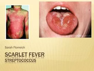
4.Erythema Marginatum • Occur in <5%. • Unique,transient,serpiginous-looking lesions of 1-2 inches in size • Pale center with red irregular margin • More on trunks & limbs & non-itchy • Worsens with application of heat • Often associated with chronic carditis
5.Subcutaneous nodules • Occur in 10% • Painless,pea-sized,palpable nodules • Mainly over extensor surfaces of joints,spine,scapulae & scalp • Associated with strong seropositivity • Always associated with severe carditis
Rheumatic Fever: Clinical Features Polyarthritis– w/ low grade fever, large joints, ( > 75%) migratory - affects 1 at a time, no permanent dysfx. Carditis- pericarditis, cardiomeagly, or valvulitis ( ~ 50%) (valvulitis is the most serious manifestation.) Chorea – late occurrence, 3 - 4 months after ( ~ 10%) infection, self-limiting, resolves in 1- 3 months. Erythema Marginatum–“classic” truncal rash, ( ~ 10%) migratory - appears & disappears within hours. (pink rash – irregular red edges – clear center) Subcutaneous Nodules– occurs late ( months (1 - 2%) after infection), painless small nodules over bony prominences - elbows, knees, spine.
Other features (Minor features) • Fever-(upto 38.3 degree centigrade) • Arthralgia • Pallor • Anorexia • Loss of weight
Jones clinical Criteria of Diagnosis: Major Criteria Migratory Polyarthritis Carditis Subcutaneous nodules Erythema marginatum Sydenham Chorea Minor Criteria Nonspecific symptoms Fever Arthralgia High ESR / CRP Prolonged PR Interval Positive:2 Major or 1 major + 2 minor Following Group-A strep. pharyngitis.
Heart Disease • Rheumatic Heart Disease – usually occurs years after initial attack. Mitral valve is more commonly involved than aortic valve. Classically, pts have mitral stenosis as a result of calcification.
Laboratory Investigations: No specific laboratory investigations* Throat culture-GABH streptococci - Cultures are usually negative. High ESR Anemia, leucocytosis Elevated C-reactive protien ASO titre >200 Todd units. (Peak value attained at 3 weeks,then comes down to normal by 6 weeks) High Anti-DNAse B titres High Acute phase reactants – CRP, SAP, Complements, Coagulation Proteins.
Laboratory Findings (Contd) • ECG- prolonged PR interval, 2nd or 3rd degree blocks,ST depression, T inversion • 2D Echo cardiography- valve edema,mitral regurgitation, LA & LV dilatation,pericardial effusion,decreased contractility
Diagnosis • Evidence of recent streptococcal infection can include: • Increased antistreptolysin O or other streptococcal antibodies (anti-DNAse B) • Positive throat culture for Group A beta-hemolytic streptococci • Positive rapid direct Group A strep test • Recent scarlet fever • Rheumatic fever is mainly a clinical diagnosis • No single diagnostic sign or specific laboratory test available for diagnosis • Diagnosis based on MODIFIED JONES CRITERIA
Differential Diagnosis • Juvenile rheumatiod arthritis • Septic arthritis • Sickle-cell arthropathy • Kawasaki disease • Myocarditis • Scarlet fever • Leukemia
Treatment • Step I- primary prevention(eradication of streptococci) • Step II- anti inflammatory treatment(aspirin,steroids) • Step III- supportive management & management of complications • Step IV- secondary prevention (prevention of recurrent attacks)
Primary prophylaxis • Timely diagnosis of GAS pharyngitis and appropriate treatment. Treatment of choice is still Penicillin as all GAS is susceptible. • Treatment administered within 10 days of onset of illness has been shown to prevent ARF. • Alternatives – amoxicillin, erythromycin, 1st generation cephalosporin
STEP I: Primary Prevention of Rheumatic Fever (Treatment of Streptococcal Tonsillopharyngitis) Agent Dose Mode Duration Benzathine penicillin G 600 000 U for patients Intramuscular Once 27 kg (60 lb) 1 200 000 U for patients >27 kg or Penicillin V Children: 250 mg 2-3 times daily Oral 10 d (phenoxymethyl penicillin) Adolescents and adults: 500 mg 2-3 times daily For individuals allergic to penicillin Erythromycin: 20-40 mg/kg/d 2-4 times daily Oral 10 d Estolate (maximum 1 g/d) or Ethylsuccinate 40 mg/kg/d 2-4 times daily Oral 10 d (maximum 1 g/d)
Step II:Anti inflammatory treatment Clinical condition Drugs
Bed rest Treatment of congestive cardiac failure: -digitalis,diuretics Treatment of chorea: -diazepam or haloperidol Rest to joints & supportive splinting 3.Step III: Supportive management & management of complications
Secondary prophylaxis • Pts diagnosed with ARF need to undergo secondary prophylaxis to prevent relapses. • Prophylaxis regimens include oral Pen VK BID, Pen G IM qmonth, oral sulfisoxazole qday, or oral erythromycin BID.
STEP IV : Secondary Prevention of Rheumatic Fever (Prevention of Recurrent Attacks) Agent Dose Mode Benzathine penicillin G 1 200 000 U every 4 weeks* Intramuscular or Penicillin V 250 mg twice daily Oral or Sulfadiazine 0.5 g once daily for patients 27 kg Oral 1.0 g once daily for patients >27 kg For individuals allergic to penicillin and sulfadiazine Erythromycin 250 mg twice daily Oral *In high-risk situations, administration every 3 weeks is justified and recommended
Duration of Secondary Rheumatic Fever Prophylaxis Category Duration Rheumatic fever with carditis and At least 10 y since last residual heart disease episode and at least until (persistent valvar disease*) age 40 y, sometimes lifelong prophylaxis Rheumatic fever with carditis 10 y or well into adulthood, but no residual heart disease whichever is longer (no valvar disease*) Rheumatic fever without carditis 5 y or until age 21 y, whichever is longer *Clinical or echocardiographic evidence.
Prognosis • Rheumatic fever can recur whenever the individual experience new GABH streptococcal infection,if not on prophylactic medicines • Good prognosis for older age group & if no carditis during the initial attack • Bad prognosis for younger children & those with carditis with valvar lesions
Assessment of CVS & Murmurs
Marfan Syndrome Tall, long extremities Associated with: aortic root dilitation, MV prolapse Acromegaly Large stature, coarse facial features, “spade” hands Associated with: Cardiac hypertrophy Turner Syndrome Web neck, hypertelorism, short stature Associated with: Aortic coarctation, pulmonary stenosis Pickwickian Syndrome Severe obesity, somnolence Associated with: Pulmonary hypertension Fredreich ataxia Lurching gait, hammertoe, pes cavus Associated with: hypertrophic cardiomyopathy Duchenne muscular dystrophy Pseudohypertrophy of the calves Cardiomyopathy Ankylosing spondylitis Straight back syndrome, stiff (“poker”) spine Associated with: AI, CHB (rare) Lentigines (LEOPARD syndrome) Brown skin macules that do not increase with sunlight Associated with: HOCM, PS General Appearance
Hereditary hemorrhagic telangiectasia (Osler-Weber-Rendu) Small capillary hemangiomas on the face or mouth Associated with: Pulmonary arteriovenous fistula Lupus Butterfly rash on face, Raynaud phenomenon- hands, Livedo reticularis Associated with: Verrucous endocarditis, Myocarditis, Pericarditis Pheochromocytoma Pale diaphoretic skin, neurofibromatosis- café-au-lait spots Associated with: Catecholamine-induced secondary dilated CM Sarcoidosis Cutaneous nodules, erythema nodosum Associated with: Secondary cardiomyopathy, heart block Tuberous Sclerosis Angiofibromas (face; adenoma sebaceum) Associated with: Rhabdomyoma Myxedema Coarse, dry skin, thinning of lateral eyebrows, hoarseness of voice Associated with: Pericardial effusion, LV dysfunction General Appearance- 2
Pathophysiology Stenosis- narrowed valve, sloews forward blood flow increases afterload,dec. CO Regurgitation (insufficiency) increases preload heart pumps same blood again blood volume and pressures reduced in front of affected valve; increased behind affected valve results in heart failure
All valvular diseases have characteristic murmurs • Damaged valve disrupts blood flow=turbulence & sound! • Caused by • Rheumatic Heart Disease • Acute conditions (infective endocarditis) • Acute MI • Congenital Heart Defects • Aging, etc
Auscultation • Use the diaphragm for high pitched sounds and murmurs • Use the bell for low pitched sounds and murmurs • Sequence of auscultation • upper right sternal border (URSB) • upper left sternal border (ULSB) • lower left sternal border (LLSB) • apex • apex - left lateral decubitus position • lower left sternal border (LLSB)- sitting, leaning forward, held expiration
Grading the Intensity of Murmurs • Grade 1 • Murmur heard with stethoscope, but not at first • Grade 2 • Faint murmur heard with stethoscope on chest wall • Grade 3 • Murmur hears with stethoscope on chest wall, louder than grade 2 but without a thrill • Grade 4 • Murmur associated with a thrill • Grade 5 • Murmur heard with just the rim held against the chest • Grade 6 • Murmur heard with the stethoscope held away and in from the chest wall
Cardiac Murmurs • Most mid systolic murmurs of grade 2/6 intensity or less are benign • Associated with physiologic increases in blood velocity: • Pregnancy • Elderly • In contrast, the following murmurs are usually pathologic: • Systolic murmurs grade 3/6 or greater in intensity • Continuous murmurs • Any diastolic murmur
Diagnostic Testing • ECHOCARDIOGRAM • Exercise testing • To assess the clinical severity of valvular heart disease • Those with inconsistent resting hemodynamics • Equivocal history of symptoms • Exercise testing in AS patients • Should be ended promptly if: • Cardiac symptoms provoked • Decrease or minimal increase (<20 mmHg) in blood pressure • Prior history of angina, congestive heart failure, or exertional syncope absolute contraindications to exercise testing • Cardiac catheterization • Usually not needed for primary evaluation
Innocent MurmursCommon in asymptomatic adults • Characterized by • Grade I – II @ LSB • Systolic ejection pattern - no with Valsalva • Normal precordium, apex, S1 • Normal intensity & splitting of second sound (S2) • No other abnormal sounds or murmurs • No evidence of LVH S1 S2