Download

1 / 40
400 likes | 479 Views
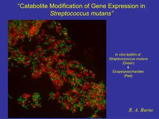
The lac repressor bound to operator sequences and the CAP-cAMP in complex with its 30 bp binding site . The TATA box and -35 region of the promoter are also indicated. Catabolite repression happens when glucose (a catabolite) levels are high . Then cyclic AMP is inhibited from forming.

E N D
Thelac repressor bound to operator sequences and the CAP-cAMP in complex with its30 bp binding site. The TATA box and -35 region of the promoter are also indicated.
Catabolite repression happens when glucose (a catabolite) levels are high. • Then cyclic AMP is inhibited from forming. • When glucose levels drop, more cAMP forms. • cAMP binds to a protein called CAP (catabolite activator protein), which is then activated to bind to the CAP binding site. • This activates transcription, perhaps by increasing the affinity of the site for RNA polymerase. • This phenomenon is called catabolite repression,
Suggested readings on regulation/dna bp Voet pp 1237-1253 Problems 2, 4 Here’s a quiz on the lac operon: http://www.bio.davidson.edu/courses/movies.html
Figure 31-39 A genetic map of the E. coli trp operon indicating the enzymes it specifies and the reactions they catalyze. Page 1251
Figure 31-40 The base sequence of the trp operator. The nearly palindromic sequence is boxed and its –10 region is overscored. Page 1251
Figure 31-41 The alternative secondary structures of trpL mRNA. Page 1252
Figure 31-42a Attenuation in the trp operon. (a) When tryptophanyl–tRNATrp is abundant, the ribosome translates trpL mRNA. Page 1253
Figure 31-42b Attenuation in the trp operon. (b) When tryptophanyl–tRNATrp is scarce, the ribosome stalls on the tandem Trp codons of segment 1.
Table 31-3 Amino Acid Sequences of Some Leader Peptides in Operons Subject to Attentuation.
Thus endeth the material for exam 1…Friday, in class The pleasure of your company is required.
Eukaryotic RNA gets a 5’ cap...
Figure 31-43The structure of the 5¢ cap of eukaryotic mRNAs. Page 1255
Figure 31-46 An electron micrograph and its interpretive drawing of a hybrid between the antisense strand of the chicken ovalbumin gene and its corresponding mRNA.
Introns in histone genes: 0!!! from Genomes
Figure 31-47 The sequence of steps in the production of mature eukaryotic mRNA as shown for the chicken ovalbumin gene. Page 1258
Figure 31-48 The consensus sequence at the exon–intron junctions of vertebrate pre-mRNAs. Page 1258
Figure 31-49 The sequence of transesterification reactions that splice together the exons of eukaryotic pre-mRNAs. 1. The 2’-OH group of a specific intron A residue in the intron nucelophilically attacks the 5’-phosphate at the 5’-intron boundary producing a 2’,5’-cyclic structure. Page 1259
2. The liberated 3’-OH group attacks the 5’-phosphate of the 5’-terminal residue of the 3’exon, forming a 3’,5’-PD bond and displacing the intron lariat.
Base pairing between U1 snRNA and the 5’ splice site of an mRNA precursor is necessary but not sufficient for splicing to occur.
The U6 snRNP associates with the 5’-end of the intron by base pairing prior to lariat formation. U6 also associates with U2 (not the band). U2 base-pairs with the conserved sequence at the splice branch point and with U6.
Splicing movie http://vcell.ndsu.nodak.edu/animations/mrnasplicing/movie.htm http://www.sumanasinc.com/webcontent/animations/content/mRNAsplicing.html Alternative splicing http://www.exonhit.com/UserFiles/Image/epissage.swf
Table 31-4 Types of Introns. Page 1259