Download
1 / 32
320 likes | 328 Views
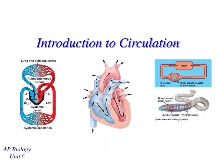
Introduction to Circulation. AP Biology Unit 6. Invertebrates with Gastrovascular Cavities. Don’t have a true circulatory system Material exchange (gases, nutrients, wastes) with the environment occurs through diffusion Why is diffusion effective here?

E N D
Introduction to Circulation AP Biology Unit 6
Invertebrates with Gastrovascular Cavities • Don’t have a true circulatory system • Material exchange (gases, nutrients, wastes) with the environment occurs through diffusion • Why is diffusion effective here? • The animals are only a few cell layers thick– materials don’t have to go across too many layers • Example: Cnidarians Slide 2 of 32
True Circulatory Systems • 3 main components in a true circulatory system: • Circulatory fluid (blood) • Tubes to transport fluid (blood vessels) • Muscular pump (heart) Slide 3 of 32
True Circulatory Systems • Blood pressure keeps the circulatory fluid moving through the system (in addition to other forces) • Blood pressure = force exerted on the walls of the blood vessels by the blood (caused primarily by the pumping of the heart) Slide 4 of 32
True Circulatory Systems • In general, higher metabolism means a more complex circulatory system • An animal either has an open or a closed circulatory system Slide 5 of 32
Open Circulatory Systems • Blood and interstitial fluid are the same (hemolymph) • Low blood pressure (less energy to circulate fluid) • Simple system of tubes • sinuses = spaces between organs • ostia = tubes that open to the body environment Slide 6 of 32
Open Circulatory System • The heart helps pump hemolymph around • Hemolymph will also be pushed back into the ostia as the animal moves around Slide 7 of 32
Closed Circulatory System • Blood is confined to tubes, so it is different from interstitial fluid • Molecules diffuse between blood and interstitial fluid • High blood pressure Slide 8 of 32
Question… • Why would higher blood pressure be beneficial? • Can get blood to areas that need it more efficiently • Allows the organism to be more active Slide 9 of 32
Closed Circulatory System • Complex system of tubes • arteries = vessels that carry blood from heart to capillaries (throughout body) • veins = vessels that carry blood from capillaries to heart (in general) • capillaries = tiny, porous vessels through which molecules diffuse in / out (throughout body) Slide 10 of 32
General Circulatory Pathway • Heart artery capillaries vein back to heart Slide 11 of 32
Comparison of Vertebrate Circulation- Fish • Gas exchange with the environment occurs in the gills • Blood pressure is highest in the artery leaving the heart to go to the lungs. Slide 12 of 32
Comparison of Vertebrate Circulation- Fish • Blood in the heart is separated (oxygenated and de-oxygenated blood are not mixed together) • Single circulation = blood goes to the heart once (continues on to the body without returning after the lungs) • 2 chambers in heart (1 atrium, 1 ventricle) Slide 13 of 32
Comparison of Vertebrate Circulation- Amphibian • Gases are exchanged with the environment in the lungs and across the skin • Blood pressure is highest where blood leaves the heart Slide 14 of 32
Comparison of Vertebrate Circulation- Amphibian • Blood in the heart is mixed– deoxygenated and newly oxygenated blood mix together in ventricle • Double circulation = blood is pumped two times from the heart– goes to the lungs, then comes back to get pumped to the rest of the body • 3 chambers in heart (2 atria, 1 ventricle) Slide 15 of 32
Comparison of Vertebrate Circulation- Reptile • Gas exchange occurs in the lungs • Blood pressure is where blood is leaving the heart Slide 16 of 32
Comparison of Vertebrate Circulation- Reptile • Blood in the heart is mixed-- deoxygenated and newly oxygenated blood mix together in partially separated ventricle • Double circulation • 3 ½ chambers in heart (2 atria, one partially separated ventricle) • Only crocodiles have fully separated ventricles Slide 17 of 32
Question… • What is the benefit of having double circulation (compared to single circulation)? • Blood can reach tissues more efficiently High blood pressure\ • This allows the organism to be more active Slide 18 of 32
Reptile Circulation • Reptiles also have a 2nd aorta • Benefit? • Can bypass the lungs when underwater (no point in sending blood to the lungs if there can’t get O2 from them) • Blood continues to flow to the body tissues (so they can still get some O2) higher activity Slide 19 of 32
Comparison of Vertebrate Circulation- Mammal & Bird • Gas Exchange occurs in the lungs • Blood pressure is where blood is leaving the heart • Blood is separated – deoxygenated and newly oxygenated blood do not mix (held in separate chambers) Slide 20 of 32
Comparison of Vertebrate Circulation- Mammal & Bird • Double circulation • 4 chambers in heart (2 atria, 2 ventricles) Slide 21 of 32
Question… • Why is having separated (compared to mixed blood) an advantage? • If blood is mixed, then deoxygenated blood that hasn’t gone to the lungs will also return to the body • Separated blood means that the blood returning to the body is all fully re-oxygenated Slide 22 of 32
Pressure and Metabolism • The inability to maintain pressure over a distance yields lower metabolism. • Pressure decreases as blood flows through tiny capillaries • Which organism can have the highest metabolic rate? • Mammals and birds (in general) Slide 23 of 32
Mammalian Heart Right side Left side • 4 chambered heart (2 atria, 2 ventricles) • Valves = flaps that keep chambers of the heart closed at the right time • Valves are needed to build pressure in heart and prevent back-flow of blood. Slide 24 of 32
Atrioventricular (AV) Valves • Located between the atria and ventricles • Tricuspid Valve • Between the right atrium and right ventricle • 3 flaps • Bicuspid (Mitral) valve • Between the left atrium and left ventricle • 2 flaps Slide 25 of 32
Semilunar valves • located at two exits for the heart • Between the right ventricle and the pulmonary artery (to lungs) • Between the left ventricle and the aorta (to the body) Slide 26 of 32
Pathway of Blood • Do you remember the pathway of blood through the body and the heart? • Use these terms: Right Atrium, Left Atrium, Right Ventricle, Left Ventricle, Pulmonary Artery, Pulmonary Vein, Aorta, Lung Capillaries, Capillaries in Top or Bottom of Body, Anterior / Posterior Vena Cava • Start where the blood first leaves the heart to go to the body Slide 27 of 32
Pathway of blood • Aorta arteries capillaries in body veins vena cava right atrium right ventricle pulmonary artery lung capillaries pulmonary vein left atrium left ventricle aorta Slide 28 of 32
Questions… • Where does the blood have the highest O2 concentration? • Just after leaving lungs (where it picked up O2) • Where does the blood have the highest CO2 concentration? • Just before getting to the lungs (hasn’t dropped off the CO2 waste yet) Slide 29 of 32
Heartbeat • The heart beat is controlled by electrical signals generated in specific cells in the heart = self excitation • Sinoatrial (SA) node = a group of specialized cells that initiates the heartbeat • Also called the pacemaker of the heart • generates electrical impulses that cause both atria to contract Slide 30 of 32
Heartbeat • Atrioventricular (AV) node • When it receives the signals from the SA node, it transfers the signals to the Bundle of His • Bundle of His spreads the signal to the Purkinje fibers in the ventricles both ventricles contract • Pathway: SA AV Bundle of His Purkinje Slide 31 of 32