Download
1 / 32
320 likes | 486 Views
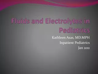
Basics of fluid therapy Zsolt Molnár 2009. Physiology. The debt…. DO 2 = (SV • P) • (Hb • 1.39 • SaO 2 +0.003 • PaO 2 ) ~ 1000ml/m (SaO 2 =100%) VO 2 = CO • (CaO 2 - CvO 2 ) ~ 250 ml/m (ScvO 2 ~70-75%). CO. CaO 2. The debt….

E N D
The debt… • DO2= (SV•P) • (Hb•1.39•SaO2+0.003•PaO2) ~ 1000ml/m (SaO2=100%) • VO2 = CO • (CaO2 - CvO2) ~ 250 ml/m (ScvO2~70-75%) CO CaO2
The debt… • DO2= (SV•P) • (Hb•1.39•SaO2+0.003•PaO2) ~ 1000ml/m (SaO2=100%) • VO2 = CO • (CaO2 - CvO2) ~ 250 ml/m (ScvO2~70-75%) • A hypovolémiás, vérző beteg: • Sokk = VO2>DO2 CO CaO2 VO2 DO2
Fluid compartments TBW ~ 40L I. c. E.c. 0.6xTBV ~ 20L I.st.~15L I.v.~5L 1/1 Coll 3/4 1/4 NaCl 4/8 3/8 1/8 5%D
Main considerations • Distribution: • Water (5%D) inthe TBW (1/8) • Na+inthee.c. (1/4) • Colloidinthei.v (1/1) • Therefore: • 1 L bloodlosscan be replacedwith… • …4 L isotonicsaline, or… • …1 L colloid. Molnár ‘99
Infusions Molnár ‘99
Clinical signs of hypovolaemia • Pulse - MAP • Capillary refill • Hourly urine output • Core – peripheral temperature differance • Moderate bleeding • Sensitivity: 20-30 % • McGee S, et al. JAMA 1999; 282: 720 Molnár ‘99
Start with a Subjective Assessment of Skin Temperature to Identify Hypoperfusion in Intensive Care Unit Patients Kaplan LJ, et al. J Trauma 2001; 50: 620-7 Cold hands = Hypoperfusion: 39% pos. pred. Cold hand + low HCO3 = Hypoperfusion: 98% pos. pred. Molnár ‘99
Peripheral lines • Features • 24 ….14 G • Color coded • Pink: 20 G • Green: 18G • White: 17G • Grey: 16G • Orange: 14G • Simple, fast • Little complications Molnár ‘99
Hagen-Poiseuille’s law • Importance: • Intravenous fluid replacement • Airways • Effective: short &thick Molnár ‘99
Peripheral lines Molnár ‘99
Central lines • Int. jugular vein • Close to the skin, „far” from the lungs • Carotid artery can be compressed • Subclavian vein • Close to the lungs, far from the skin • Subcl. artery cannot be compressed • Femoral vein • Far fromthe skin, close to the groin • Fem. artery can be compressed Molnár ‘99
CVC: complication • Pain • Use local anaesthesia all the time • Arterial puncture • Inc ase of clotting disorder – use femoral, jugular approach • Pneumothorax • Subclavian > int. jugular • Catheter infection • Femoral > int. jugular > subclavian • Prevention: regular (7-10 days) change Molnár ‘99
CVC catheter set Molnár ‘99
Seldinger’s technique Molnár ‘99
US guided puncture Molnár ‘99
CVC in the int. jugular vein Molnár ‘99
CVC in the subclavian vein Molnár ‘99
Position of the tip of the catheter Molnár ‘99
Mortality Choi PT et al. Crit Care Med 1999; 27: 200
SAFE Finfer S et al. SAFE study. N Eng J Med 2004; 350: 2247
SAFE Finfer S et al. SAFE study. N Eng J Med 2004; 350: 2247
Instead of summary • „Early Goal-Directed Therapy” (EGDT) Rivers E et al. N Engl J Med 2001; 345: 1368 • Septic patients treated on A&E for 6 hours: • Control group (n=133): • O2 • CVP: 8-12 mmHg • MAP >65 mmHg • EGDT group (n=130): • Same as above • ScvO2 > 70% • More fluid, blood • More dobutamine Mortality: 46 vs. 30% (p=0.009)
Haemodinamics • Otto Frank, Ernest Starling – 1914: „Law of the heart” • „The mechanical energy set free in the passage from the resting to the active state is a function of the length of the fiber„ • „Within physiological limits, the force of contraction is directly proportional to the initial length of the muscle fiber” • Most common reasons of HF: • Reduced circulating volume • Reduced pump function SV „End point” of resusscitation EDV Starling EH. The Linacre Lecture on the Law of the Heart. London; 1918 Starling EH. J R Army Med Corps. 1920; 34: 258-262 Molnár ‘99
Summary • Basic physiological knowledge • Read the label! • Fluid therapy is also revolving around: O2 Diagnosis can wait but cells can’t!