Download
1 / 1
10 likes | 163 Views
Imaging HIFU Lesions Using Ultrasound Andrew Draudt and Robin Cleveland Department of Aerospace and Mechanical Engineering, Boston University, Boston, MA 02215. R2. Fundamental Science. R1. R3. Validating TestBEDs. Bio-Med. Enviro-Civil. S2. S3. S4. S1. S5.

E N D
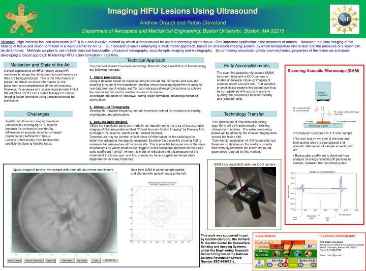
Imaging HIFU Lesions Using Ultrasound Andrew Draudt and Robin Cleveland Department of Aerospace and Mechanical Engineering, Boston University, Boston, MA 02215 R2 FundamentalScience R1 R3 ValidatingTestBEDs Bio-Med Enviro-Civil S2 S3 S4 S1 S5 SAM transducer (left) with new CCD camera Optical image of bovine liver sample with 2mm dia. burn from hot filament. Data from SAM of same sample,scaled and aligned with optical image to the left 3-Level Diagram PI CONTACT INFORMATION Prof. Robin Cleveland Aerospace and Mechanical Engineering Dept. Boston University, Boston, MA, 02215 Phone: 617-353-7767 Email: robinc@bu.edu Abstract: High intensity focused ultrasound (HIFU) is a non-invasive method by which ultrasound can be used to thermally ablate tissue. One important application is the treatment of tumors. However, real-time imaging of the heating of tissue and lesion formation is a major barrier for HIFU. Our research involves employing a multi-modal approach, based on ultrasound imaging system, by which temperature distribution and the presence of a lesion can be determined. Methods we plan to use include classical backscatter, ultrasound tomography, acousto-optic imaging and elastography. By combining acoustical, optical and mechanical properties of the lesion we anticipate developing a robust approach to tracking HIFU lesion formation in real time. Technical Approach Motivation and State of the Art Early Accomplishments Our planned research involves improving ultrasonic image resolution of lesions using the following methods: 1. Signal processing. Using a detailed model for backscattering to include the diffraction and acoustic coupling function of the transducer, develop new processing algorithms to apply to raw data from our Anologic and Terrason ultrasound imaging machines to achieve the necessary contrast to resolve lesions in formation. Investigate the onset of “shadows” during lesion formation, indicating increased attenuation. 2. Ultrasound Tomography. Develop Born-based frequency-domain inversion method for variations in density, soundspeed and attenuation. 3. Acousto-optic imaging. Utilize the significant advances made in our department in the area of acousto-optic imaging (AOI) (see poster entitled “Pulsed Acousto-Optioc Imaging” by Puxiang Lai) to image HIFU lesions, which exhibit optical contrast. Temperature may be another critical piece of information for the radiologist to determine adequate therapeutic exposure. Examine the possibility of using AOI to measure the temperature at the lesion site. This is possible because one of the main mechanisms by which photons are “tagged” in this technique depends on the piezo-optic coefficient (“dn/dp”, where n is index of refraction and p is pressure) of the material at the focus spot, and this is known to have a significant temperature dependence for many materials. Scanning Acoustic Microscope (SAM) Clinical applications of HIFU therapy utilize MRI machines to image the ultrasound-induced lesions as they are being produced. This is the only means at present to obtain accurate information on the placement and completeness of the cell necrosis. However, it’s expense and space requirements inhibit the adoption of HIFU as a viable therapy for cancer. Imaging lesion formation using ultrasound would be preferable. The scanning acoustic microscope (SAM) has been fitted with a CCD camera to enable underwater close-up imaging of samples under acoustic test. Flat samples of small tissue regions like lesions can thus be co-registered with acoustic scans to quantify the boundaries between healthy and “cooked” cells. Focused Transducer Pf = pulse reflected off front of sample Pb = pulse reflected off back of sample Challenges Technology Transfer Traditional ultrasonic imaging has been unsuccessful at imaging HIFU lesions because it’s contrast is provided by differences in acoustic reflection strength (backscatter coefficient) in tissue. Lesions unfortunately have backscatter coefficients close to healthy issue. *The application of new data processing algorithms will be implemented on existing ultrasound machines. The extra processing power will be offset by the smaller imaging area around the lesion site. *Commercial realization of AOI is possible, but there are no devices on the market currently that remotely resemble the laser/ultrasound geometries required by this method. sample Th = sample thickness hard backing material • Transducer is scanned in X-Y over sample. • The size and arrival time of the front and back pulses give the soundspeed and acoustic attenuation of sample at each point XY. • Backscatter coefficient is obtained from analysis of energy reflected off particles in sample, between front and back pulse. Typical received waveform Amplitude (volts) Time (samples) Backscatter region Front echo Back echo This work was supported in part by Gordon-CenSSIS, the Bernard M. Gordon Center for Subsurface Sensing and Imaging Systems, under the Engineering Research Centers Program of the National Science Foundation (Award Number EEC-9986821).