Download
1 / 39
410 likes | 520 Views
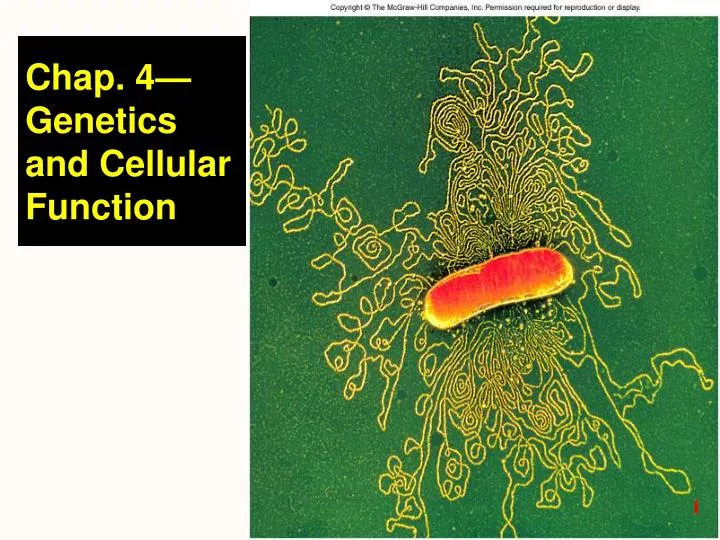
Chap. 4—Genetics and Cellular Function. Ch. 4 Study Guide. Critically read Chapter 4 up to page 129 right before 4.3 “DNA Replication and the Cell Cycle” section Comprehend Terminology (those in bold in the textbook)

E N D
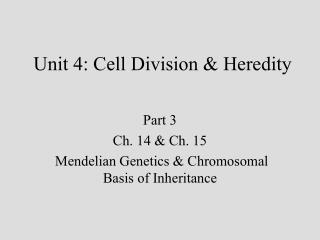
Ch. 4 Study Guide Critically read Chapter 4 up to page 129 right before 4.3 “DNA Replication and the Cell Cycle” section Comprehend Terminology (those in bold in the textbook) Study-- Figure questions, Think About It questions, and Before You Go On (section-ending) questions Do end-of-chapter questions: Testing Your Recall— 2, 4, 5, 6, 7, 18 True or False– 1, 2, 4-7
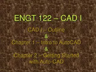
§ DNA structure (1) • General • DNA– deoxyribonucleic acid • Most human cells have 46 molecules of DNA • A uniform diameter of 2 nm and the average length @ 2-in. • Molecular level— • Nucleic acids (DNA + RNA) are polymers of __________________________ • A nucleotide consists of (1) ________ + (2) ________ + (3) ___________________ • DNA is a double helix (@ spiral staircase) Fig. 4.1 a +b and 4.2
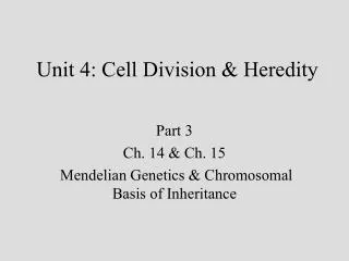
Adenine See next slide A nucleotide consists of three components NH2 C N N C HC CH C N N H O O HO P O CH2 OH H H H H OH H Phosphate Deoxyribose (a)
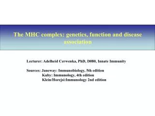
Five nitrogenous bases: O N Purines NH2 C H N C C CH C C NH HN C NH N C C N N C NH2 H Adenine (A) Guanine (G) Pyrimidines NH2 CH3 O H C C C C H C N HC NH N C N C H H O O T: Only in DNA Cytosine (C) Thymine (T) O C H N C H C C H N O H Uracil (U) U: Only in RNA (b)
§ DNA Structure (2) Space-filling model “Twisted ladder”
§ DNA Structure (3) • DNA = a double helix molecule; a spiral staircase; a soft rubber ladder that you can twist • Details: • Each sidepiece is a backbone-- composed of phosphate groups alternating with the sugar deoxyribose. • Step-like connections-- between the backbones are pairs of nitrogenous bases. • The arrangement of these nitrogenous bases– How? (Next slide)
§ DNA Structure (4) Sugar-phosphate backbone Sugar-phosphate backbone Law of complementary base pairing: • Base pairs (2 kinds): • A-T and C-G • Nitrogenous bases form hydrogen bonds Segment of DNA
§ DNA Function • Carry instructions of genes for protein synthesis • A gene – a segment of DNA that codes for one polypeptide (or closely related proteins) • Genes determine the characteristics of a species and each individual • Genome - all the genes of one person • humans have estimated 25,000-35,000 genes (2% of DNA) • The other 98% of DNA is noncoding – either “junk” or organizational DNA
Think About It • What would be the base sequence of the DNA strand across from ATTGACTCG? • If a DNA molecule were known to be 20% adenine, predict its percentage of cytosine.
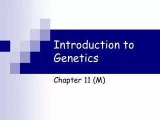
§ Chromatin and Chromosomes • Chromatin—filamentous material making up 46 chromosomes (DNA and proteins) in the interphase nucleus • Chromatin appears like “beads on a string” packed together (Fig. 4.2 a-f) • The beaded string is divided into segments called nucleosomes (consist of histones and linker DNA) • In dividing cells, DNA coils and supercoils itself to form chromosomes (can be seen with light microscope) . Fig. 4.5
2 nm 1 DNA double helix
Core particle Linker DNA 11 nm 2 DNA winds around core particles Nucleosome
30 nm 3 Nucleosomes Fold into zigzag fiber
4 300 nm fiber is thrown into irregular loops
In dividing cells only 5 looped chromatin coils further into a chromatid 700 nm
Chromatids Centromere 700 nm 6 Chromosome at the midpoint (metaphase) of cell division
Chromosome structure at metaphase Kinetochore Centromere Sister chromatids (a)
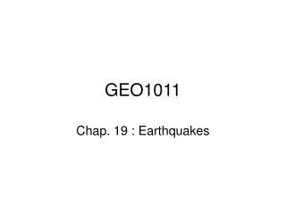
§ RNA (ribonucleic acids): Structure • RNA-- much smaller than DNA (fewer bases) • messenger RNA (mRNA) has over 10,000 bases • ribosomal RNA (rRNA) • transfer RNA (tRNA), smallest, has 70 - 90 bases (Fig. 4.8) • Are these bases (of RNAs) paired or unpaired? • Onlyonenucleotide chain (not a double helix) • ribose replaces deoxyribose as the sugar • uracil replaces thymine as a nitrogenous base
Figure 4.8 Transfer RNA (tRNA)
§ RNA: Function • DNA directs the synthesis of proteins by means of its smaller cousins, the RNAs • Essential function of RNA-- • interpretDNA code • directprotein synthesis in the cytoplasm • (Location) RNA works mainly in the cytoplasm while DNA remains safely behind in the nucleus • Table 4.1 is an excellent summary (Comparison of DNA/RNA)
Check Point Questions What are four nitrogenous bases found in RNA? a) U, G, C, T; b) A, G, C, T c) A, U, G, C; d) A, T, G, C In RNA, when does the secondary structure called a hairpin form? When hydrophilic residues act with water When complementary base pairing between ribonucleotides on the same strand creates a stem-and-loop structure When complementary base pairing forms a double helix
§ Protein Synthesis: Genetic Control of Cell Action • DNA codes for the synthesis of all cell proteins • including enzymes that direct the synthesis of nonproteins • For example, Testosterone production • Different cells synthesize different proteins • Why? • Due to differing gene activation • See Fig. 4.13 (next slide)
§ Summary of Protein Synthesis • DNA contains a genetic code that specifies which proteins a cell can make; protein synthesis as: DNA mRNA protein • Transcription (DNA mRNA); What? Details? • messenger RNA (mRNA) is formed next to an activated gene • mRNA migrates to cytoplasm • Translation (mRNA protein) (Fig. 4.7) What? How? • mRNA code is “read” by ribosomes • transfer RNA (tRNA) delivers the amino acids to the ribosome • Ribosomes assemble amino acids in the order . . .
§ Genetic Code • Def. -- System that enables the 4 nucleotides (A,T,G,C) to code for the 20 amino acids • Base triplet: (of DNA) Fig. 4.10 • Def.– A sequence of 3 nucleotides that stand for 1 amino acid • found on DNA molecule (ex. TAC codes for AUG in mRNA) • Codon: (genetic code is expressed in terms of codons) • Def.--“mirror-image” sequence of nucleotides found in mRNA (ex. AUG is the codon of mRNA, code for methionine, an amino acid) (Table 4.2) • 64 possible codons (43) • often 2-3 codons represent the same amino acid • start codon = AUG • 3 stop codons = UAG, UGA, UAA
A Seven base triplets B
§ Protein Synthesis (details) • Three sites of the large subunit of ribosome: • P (peptidyl) site— • A (acceptor) site— • E (exit) site--
Watch a video-- An animation: protein synthesis, when available