Download
1 / 24
240 likes | 383 Views
Muscles and Movement:. Body Structure 37.3 Muscular System. Muscles and Movement: Every time you move, you use muscles. Walking and running require precisely timed and controlled contractions of many skeletal muscles working in a coordination.

E N D
Muscles and Movement: Body Structure 37.3 Muscular System
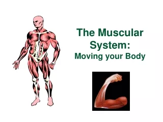
Muscles and Movement: Every time you move, you use muscles. Walking and running require precisely timed and controlled contractions of many skeletal muscles working in a coordination. Even when you are idle, many of your muscles, including those in your neck and back, remain partially contracted to maintain your balance and posture. Body Structure 37.3 Muscular System
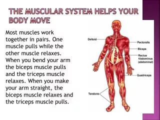
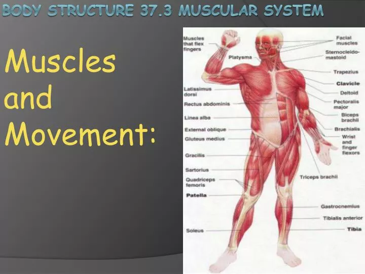
Movement of the Skeleton: Muscles can move body parts because muscles are attached to bones by strips of dense connective tissue called tendons. One attachment of the muscle, the origin, is a bone that remains stationary during a muscle contraction. The muscle pulls against the origin. The other attachment, the insertion, is the bone that moves when the muscle contracts. Movement occurs when a muscle contraction pulls the muscle’s insertion toward its origin. Movement of the Skeleton
Skeletal muscles are generally attached to the skeleton in opposing pairs. One muscle in a pair pulls a bone in one direction, and the other pulls the bone in an opposite direction. In the limbs, each opposing pair of muscles pulls the bone in an opposite direction. In the limbs, each pair of muscles includes a flexor muscle and an extensor muscle. A flexor muscle causes a joint to bend while an extensor muscle causes a joint to straighten. Movement of the Skeleton
Muscles contain some connective tissue, which holds muscle cells together and provides elasticity. Muscle tissues also contain large amounts of contractile protein filaments. These protein filaments, called actin and myosin, enable muscles to contract. Actin and myosin are usually found in the cytoskeleton of eukaryotic cells, but they are far more abundant in muscle cells. Other characteristics of muscle tissue include the ability to stretch or expand, and the ability to respond to stimuli, such as signal molecules released by nerve cells. Muscle Structure
Skeletal muscle tissue consists of many parallel elongated cells called muscle fibers. Each muscle fiber contains small cylindrical structures called myofibrils. Myofibrils have alternating light and dark bands that produce a characteristic striated, or striped, appearance when viewed under a microscope. Muscle Structure
In the center of each light band is a structure called a Z line, which anchors actin filaments. The area between two Z lines is called a sarcomere. Thus, a myofibril is a grouping of sarcomeres linked end to end. Each sarcomere contains thin and thick protein filaments that move and interact with each other. The thin filaments are actin, and the thick filaments are myosin. The filaments run parallel to one another along the length of the sacromere. The dark bands that occur in the middle of the sarcomere are regions where the thick filaments and the thin filaments overlap. Muscle Structure
Muscle Contraction: Muscle contraction occurs in the sarcomeres of myofibrils. The overlapping arrangement of the thick and thin protein filaments in a sarcomere enables muscle contraction. Muscle Contraction happens in three steps Before a muscle is stimulated, the sarcomere is relaxed. Myosin and actin filaments partially overlap one another. Muscle Contraction
2: A muscle contraction usually begins when a muscle fiber is stimulated by signal molecules released by a nerve cell. This causes myosin and actin filaments to “slide” along one another so that they overlap even more. The sarcomere becomes shorter as the Z lines are pulled closer together. Muscle Contraction
3: The sarcomere is fully contracted, and myosin and actin completely overlap one another. This shortening of sarcomeres occurs down the entire length of the muscle fiber. Muscle contraction
What determines the force of a contraction? A muscle exerts the greatest force when all of it’s fibers are contracted. When a fiber is stimulated, it’s sarcomeres contract. The total amount of force a muscle exerts depends on how often fibers are stimulated and how many muscle fibers contract at once. Muscle Contraction
How is the force of muscle contraction controlled? As different numbers of fibers in a muscle contract at one time, the total force exerted by the total contraction varies. For example, the total amount of force needed to lift a pencil is much less force than what is needed to lift a brick. Muscle Contraction:
The set of muscle fibers activated by a nerve cell is called a motor unit. Every time a nerve cell activates it’s motor unit, all the fibers in that unit contract. Muscles that require a fine degree of control , such as muscles that move the fingers, have only a few muscle fibers in each unit. Large muscles, such as the muscles in the leg, have several hundred muscle fibers in each motor unit. Muscle Contraction:
How do actin and myosin cause sarcomeres to shorten? Myosin filaments have long, finger-like projections with an enlarged “head” at one end. Actin filaments contain many sites to which myosin can bind during a muscle contraction. Stimulation of a muscle fiber leads to the exposure of these binding sites on actin filaments. The myosin heads attach to binding sites on actin filaments, causing myosin to move relative to actin. Interaction of Myosin and Actin
Step 1: Muscle Contraction begins as a myosin head attaches to an exposed binding site on an actin filament Interaction of Myosin and Actin
Step 2: The Myosin head rotates, causing the actin filament to “slide” against the myosin filament. This sliding causes the filaments to overlap one another Interaction of Myosin and Actin
Step 3: ATP is used as the myosin head detaches and snaps back into it’s original binding position. The myosin head reattaches to actin at a binding site farther along the actin filament. When the myosin heads can not move farther , they release momentarily to reposition themselves to grab the actin and pull again. Interaction of Myosin and Actin
The myosin heads “walk” along the actin filaments, essentially stepping at each available binding site. This grabbing and pulling action is repeated, causing the sacromere to shorten as the Z lines are pulled closer together. Interaction of Myosin and Actin
A lot of energy is needed to power muscle contraction. ATP is used each time a myosin head moves from one binding site on an actin filament to another. Without ATP, myosin heads would remain attached to actin filaments, keeping the muscle contracted. Calcium ions are needed for binding sites to be exposed on actin filaments. Without calcium ions and ATP, a muscle would not be able to contract. ATP and Calcium Ions
The ATP used to power contractions is usually supplied by aerobic respiration. During prolonged exercise, such as long distance walking, oxygen is consumed at a sustainable steady rate and aerobic respiration yields most of the ATP. However, during brief intense activity, such as sprinting or weight lifting, anaerobic processes take over. Most of the ATP used in these activities comes from glycolysis, as the oxygen available to muscle cells becomes rapidly depleted. Aerobic and Anaerobic energy Pathways
When both anaerobic and aerobic energy pathways become insufficient for muscle contraction, muscles can only use glycogen as an energy source. When ATP consumption exceeds ATP production, muscle fatigue and soreness may result, leaving muscle fibers unable to recover from contraction. Aerobic and Anaerobic energy Pathways
Consistent aerobic exercise makes the heart pump more efficiently and thus increases the energy available to muscles as a result of improved blood circulation. More oxygen is extracted by the body with each breath, increasing the oxygen supply to muscles. More ATP is available for muscle contractions, thereby reducing muscle fatigue. The increase in muscle efficiency results in greater endurance, or the ability to continue exercising. Exercise and Fitness
Resistance exercise such as weight lifting can increase muscle size and strength. Resistance exercises are mostly anaerobic, so they do not usually improve the uptake of oxygen to muscles. Muscle mass is increased by resistance training. The amount of tension and the rate of exercise are both important factors. However, the short term demands of strength training do not cause the circulatory changes that increase endurance. Exercise and Fitness