Download
1 / 61
710 likes | 1.41k Views
Kidney Function Testing. DR. MALIK ALQUB MD. PHD. An Introduction to the Urinary System. Transports urine towards bladder. Temporarily store urine . Produces urine. Conducts urine to exterior. The Function of Urinary System. A). Excretion & Elimination:

E N D
Kidney Function Testing DR. MALIK ALQUB MD. PHD.
An Introduction to the Urinary System Transports urine towards bladder Temporarily store urine Produces urine Conducts urine to exterior
The Function of Urinary System A) • Excretion & Elimination: • removal of organic wastes products from body fluids (urea, creatinine, uric acid) • Homeostatic regulation: • Water -Salt Balance • Acid - base Balance • Enocrine function: • Hormones B) C)
The excretory function A) • excretion of excess electrolytes, nitrogenous wastes and organic acids • The maximal excretory rate is limited or established by their plasma concentrations and the rate of their filtration through the glomeruli • The maximal amount of substance excreted in urine does not exceed the amount transferred through the glomeruli by ultrafiltration except in the case of those substances capable of being secreted by the tubular cells.
B) Homeostatic Functions • Regulate blood volume and blood pressure: • by adjusting volume of water lost in urine • releasing renin from the juxtraglomerular apparatus 1) Water -Salt Balance Blood volume is associated with Salt volume. The greater the blood volume the greater the blood pressure. Removing water lowers blood pressure • Regulate plasma ion concentrations: • sodium, potassium, and chloride ions (by controlling quantities lost in urine) • calcium ion levels
B) Homeostatic Functions 2) Acid-Base Balance (Help stabilize blood pH) The kidneys control this by excreting H+ ions and reabsorbing HCO3 (bicarbonate). If plasma pH is low (acidic). H+ secretion in the urine and HCO3¯ reabsorption back to the plasma increases thusurine becomes more acidic, and the plasma more alkaline. If plasma pH is high (alkaline). H+ secretion in the urine and HCO3¯ reabsorption back to the plasma decreases thusurine becomes more alkaline, and the plasma more acidic.
C) The endocrine function • Kidneys have primary endocrine function since they produce hormones (erythropoietin, renin and prostaglandin). • Erythropoietin is secreted in response to a lowered oxygen content in the blood. It acts on bone marrow, stimulating the production of red blood cells. • Renin the primary stimuli for renin release include reduction of renal perfusion pressure and hyponatremia. Renin release is also influenced by angiotension II and ADH. • The kidneys are primarily responsible for producing vitamin D3 • In addition, the kidneys are site of degradation for hormones such as insulin and aldosterone,
KIDNEY STRUCTURE AND URINE FORMATION
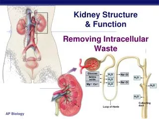
Each kidney consists of one million functional units: Nephrone Nephron structure
The Glomerulus Blood pressure inside of the glomerulus is very high. Because ofdifferences in the resistance between the afferent and efferent arterioles. Forces the fluids and some solids out of the blood into the glomerular capsule.
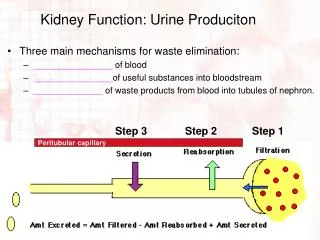
Urine Formation Urine formation requiers : a) Glomerular Filtration Due to differences in pressure water, small molecules move from the glomerulus capillaries into the glomerular capsule b) Tubular reabsorption many molecules are reabsorbedfrom the nephron into the capillary(diffusion, facilitated diffusion, osmosis, and active transport) i.e. Glucose is actively reabsorbed with transport carriers. If the carriers are overwhelmed glucose appears in the urine indicating diabetes Tubular secretion Substances are actively removed from blood and added to tubular fluid (active transport) ie. H+, creatinine, and some drugs are moved by active transport from the blood into the distal convoluted tubule c)
Urine Formation Glomerular Filtration The first step in the production of urine is called glomerular filtration. Filtration: the forcing of fluids and dissolved substances through a membrane by pressure occurs in the renal corpuscle of the kidneys across the endothelial capsular membrane (Bowman's) capsule. - The resulting fluid is called the filtrate. - Filtration is a passive process. - The total filtration rate of the kidneys is mainly determined by the difference between the blood pressure in the glomerular capillaries and the hydrostatic pressure in the lumen of the nephron
Glomerular Filtration Rate Glomerular Filtration Rate (GFR) Affected by: 1). Total filtration surface area 2). Membrane permeability 3). Net Filtration Pressure (as NFP goes up so does the GFR) The amount of filtrate that flows out of all the renal corpuscles of both kidneys every minute is called the glomerular filtration rate (GFR). In the normal adult, this rate is about 120 ml/min; about 180 liters/Day
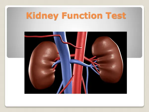
Biochemical Tests of Renal Function • Measurement of GFR • Clearance tests • Plasma creatinine • Urea, uric acid and β2-microglobulin • Renal tubular function tests • Osmolality measurements • Specific proteinurea • Glycouria • Aminoaciduria • Urinalysis • Appearance • Specific gravity and osmolality • pH • osmolality • Glucose • Protein • Urinary sediments
When should you assess renal function? • Older age • Family history of Chronic Kidney disease (CKD) • Decreased renal mass • Low birth weight • Diabetes Mellitus (DM) • Hypertension (HTN) • Autoimmune disease • Systemic infections • Urinary tract infections (UTI) • Nephrolithiasis • Obstruction to the lower urinary tract • Drug toxicity
Biochemical Tests of Renal Function • Measurement of GFR • Clearance tests • Plasma creatinine • Urea, uric acid and β2-microglobulin
Biochemical Tests of renal function In acute and chronic renal failure, there is effectively a loss of function of whole nephrons • Filtration is essential to the formation of urine tests of glomerular function are almost always required in the investigation and management of any patient with renal disease. • The most frequently used tests are those that assess either the GFR or the integrity of the glomerular filtration barrier.
Measurement of glomerular filtration rate GFR can be estimated by measuring the urinary excretion of a substance that is completely filtered from the blood by the glomeruli and it is not secreted, reabsorbed or metabolized by the renal tubules. • Clearance is defined as the (hypothetical) quantity of blood or plasma completely cleared of a substance per unit of time. • Clearance of substances that are filtered exclusively or predominantly by the glomeruli but neither reabsorbed nor secreted by other regions of the nephron can be used to measure GFR. • Inulin • The Volume of blood from which inulin is cleared or completely removed in one minute is known as the inulin clearance and is equal to the GFR. • Measurement of inulin clearance requires the infusion of inulin into the blood and is not suitable for routine clinical use V is not urine volume, it is urine flow rate
Biochemical Tests of Renal Function • Measurement of GFR • Clearance tests • Plasma creatinine • Urea, uric acid and β2-microglobulin
Creatinine • 1 to 2% of muscle creatine spontaneously converts to creatinine daily and released into body fluids at a constant rate. • Endogenous creatinine produced is proportional to muscle mass, it is a function of total muscle mass the production varies with age and sex • Dietary fluctuations of creatinine intake cause only minor variation in daily creatinine excretion of the same person. • Creatinine released into body fluids at a constant rate and its plasma levels maintained within narrow limits Creatinine clearance may be measured as an indicator of GFR.
Creatinine clearance and clinical utility • The most frequently used clearance test is based on the measurement of creatinine. • Small quantity of creatinine is reabsorbed by the tubules and other quantities are actively secreted by the renal tubules So creatinine clearance is approximately 7% greater than inulin clearance. • The difference is not significant when GFR is normal but when the GFR is low (less 10 ml/min), tubular secretion makes the major contribution to creatinine excretion and the creatinine clearance significantly overestimates the GFR.
Creatinine clearance clinical utility • An estimate of the GFR can be calculated from the creatinine content of a 24-hour urine collection, and the plasma concentration within this period. • The volume of urine is measured, urine flow rate is calculated (ml/min) and the assay for creatinine is performed on plasma and urine to obtain the concentration in mg per dl or per ml. Creatinine clearance in adults is normally about of 120 ml/min, The accurate measurement of creatinine clearance is difficult, especially in outpatients, since it is necessary to obtain a complete and accurately timed sample of urine
Creatinine clearance and clinical utility • The 'clearance' of creatinine from plasma is directly related to the GFR if: • The urine volume is collected accurately • There are no ketones or heavy proteinuria present to interfere with the creatinine determination. • It should be noted that the GFR decline with age (to a greater extent in males than in females) and this must be taken into account when interpreting results.
Use of Formulae to Predict Clearance • Formulae have been derived to predict Creatinine Clearance (CC) from Plasma creatinine. • Plasma creatinine derived from muscle mass which is related to body mass, age, sex. • Cockcroft & Gault Formula • CC = k[(140-Age) x weight(Kg))] / serum Creatinine (µmol/L) • k = 1.224 for males & 1.04 for females • Modifications required for children & obese subjects • Can be modified to use Surface area
Biochemical Tests of Renal Function • Measurement of GFR • Clearance tests • Plasma creatinine • Urea, uric acid and β2-microglobulin
Measurement of nonprotein nitrogen-containing compounds Catabolism of proteins and nucleic acids results in formation of so called nonprotein nitrogenous compounds. Protein Proteolysis, principally enzymatic Amino acids Transamination and oxidative deamination Ammonia Enzymatic synthesis in the “urea cycle” Urea
Plasma Urea Urea is the major nitrogen-containing metabolic product of protein catabolism in humans, • Its elimination in the urine represents the major route for nitrogen excretion. • More than 90% of urea is excreted through the kidneys, with losses through the GIT and skin • Urea is filtered freely by the glomeruli • Plasma urea concentration is often used as an index of renal glomerular function • Urea production is increased by a high protein intake and it is decreased in patients with a low protein intake or in patients with liver disease.
Plasma Urea • Many renal diseases with various glomerular, tubular, interstitial or vascular damage can cause an increase in plasma urea concentration. • The reference interval for serum urea of healthy adults is 5-39 mg/dl. Plasma concentrations also tend to be slightly higher in males than females. High protein diet causes significant increases in plasma urea concentrations and urinary excretion. • Measurement of plasma creatinine provides a more accurate assessment than urea because there are many factors that affect urea level. • Nonrenal factors can affect the urea level (normal adults is level 5-39 mg/dl) like: • Mild dehydration, • high protein diet, • increased protein catabolism, muscle wasting as in starvation, • reabsorption of blood proteins after a GIT haemorrhage, • treatment with cortisol or its synthetic analogous
Clinical Significance • States associated with elevated levels of urea in blood are referred to as uremia or azotemia. • Causes of urea plasma elevations: • Prerenal: renal hypoperfusion • Renal: acute tubular necrosis • Postrenal: obstruction of urinary flow
Uric acid • In human, uric acid is the major product of the catabolism of the purine nucleosides, adenosine and guanosine. • Purines are derived from catabolism of dietary nucleic acid (nucleated cells, like meat) and from degradation of endogenous nucleic acids. • Overproduction of uric acid may result from increased synthesis of purine precursors. • In humans, approximately 75% of uric acid excreted is lost in the urine; most of the reminder is secreted into the GIT
Uric acid • Renal handling of uric acid is complex and involves four sequential steps: • Glomerular filtration of virtually all the uric acid in capillary plasma entering the glomerulus. • Reabsorption in the proximal convoluted tubule of about 98 to 100% of filtered uric acid. • Subsequent secretion of uric acid into the lumen of the distal portion of the proximal tubule. • Further reabsorption in the distal tubule. • Hyperuricemia is defined by serum or plasma uric acid concentrations higher than 7.0 mg/dl (0.42mmol/L) in men or greater than 6.0 mg/dl (0.36mmol/L) in women
Plasma β2-microglobulin • β2-microglobulin is a small peptide (molecular weight 11.8 kDa), • It is present on the surface of most cells and in low concentrations in the plasma. • It is completely filtered by the glomeruli and is reabsorbed and catabolized by proximal tubular cells. • The plasma concentration of β2-microglobulin is a good index of GFR in normal people, being unaffected by diet or muscle mass. • It is increased in certain malignancies and inflammatory diseases. • Since it is normally reabsorbed and catabolized in the tubules, measurement of β2-microglobulin excretion provides a sensitive method of assessing tubular integrity.
Biochemical Tests of Renal Function • Renal tubular function tests • Osmolality measurements • Specific proteinurea • Glycouria • Aminoaciduria
Renal tubular function tests • To ensure that important constituents such as water, sodium, glucose and a.a. are not lost from the body, tubular reabsorption must be equally efficient • Compared with the GFR as an assessment of glomerualr function, there are no easily performed tests which measure tubular function in quantitative manner • Investigation of tubular function: • Osmolality measurements in plasma and urine; normal urine: plasma osmolality ratio is usually between 1.0-3.0 • Specific proteinuria • Glycosuria • Aminoaciduria
Assessment of glomerular integrity • Proteinuria may be due to: 1. An abnormality of the glomerular basement membrane. 2. Decreased tubular reabsorption of normal amounts of filtered proteins. 3. Increased plasma concentrations of free filtered proteins. 4. Decreased reabsorption and entry of protein into the tubules consequent to tubular epithelial cell damage. • Measurement of individual proteins such as β2-microglobulin have been used in the early diagnosis of tubular integrity. • With severe glomerular damage, red blood cells are detectable in the urine (haematuria), the red cells often have an abnormal morphology in glomerular disease. • Haematuria can occur as a result of lesions anywhere in the urinary tract,
Proteinuria • The glomerular basement membrane does not usually allow passage of albumin and large proteins. A small amount of albumin, usually less than 25 mg/24 hours, is found in urine. • Urinary protein excretion in the normal adult should be less than 150 mg/day. • When larger amounts, in excess of 250 mg/24 hours, are detected, significant damage to the glomerular membrane has occurred. • Quantitative urine protein measurements should always be made on complete 24-hour urine collections. • Albumin excretion in the range 25-300 mg/24 hours is termed microalbuminuria
Proteinuria • Normal < 150 mg/24h. • TYPES OF PROTEINURIA • Glomerular proteinuria • Tubular proteinuria • Overflow proteinuria
Glomerular proteinuria • Glomerular proteinuria — Glomerular proteinuria is due to increased filtration of macromolecules (such as albumin) across the glomerular capillary wall. The proteinuria associated with diabetic nephropathy and other glomerular diseases, as well as more benign causes such as orthostatic or exercise-induced proteinuria fall into this category. Most patients with benign causes of isolated proteinuria excrete less than 1 to 2 g/day
Tubular proteinuria • Low molecular weight proteins — such as ß2-microglobulin, immunoglobulin light chains, retinol-binding protein, and amino acids — have a molecular weight that is generally under 25,000 in comparison to the 69,000 molecular weight of albumin. These smaller proteins can be filtered across the glomerulus and are then almost completely reabsorbed in the proximal tubule. Interference with proximal tubular reabsorption, due to a variety of tubulointerstitial diseases or even some primary glomerular diseases, can lead to increased excretion of these smaller proteins
Overflow proteinuria • Increased excretion of low molecular weight proteins can occur with marked overproduction of a particular protein, leading to increased glomerular filtration and excretion. This is almost always due to immunoglobulin light chains in multiple myeloma, but may also be due to lysozyme (in acute myelomonocytic leukemia), myoglobin (in rhabdomyolysis), or hemoglobin (in intravascular hemolysis
Biochemical Tests of Renal Function • Urinalysis • Appearance • Specific gravity and osmolality • pH • Glucose • Protein • Urinary sediments
Urinalysis • Urinalysis is important in screening for disease is routine test for every patient, and not just for the investigation of renal diseases • Urinalysis comprises a range of analyses that are usually performed at the point of care rather than in a central laboratory. • Urinalysis is one of the commonest biochemical tests performed outside the laboratory. • Examination of a patient's urine should not be restricted to biochemical tests.
Urinalysis using disposable strips • Biochemical testing of urine involves the use of commercially available disposable strips When the strip is manually immersed in the urine specimen, the reagents react with a specific component of urine in such a way that to form color • Colour change produced is proportional to the concentration of the component being tested for. • To test a urine sample: • fresh urine is collected into a clean dry container • the sample is not centrifuged • the disposable strip is briefly immersed in the urine specimen; • The colour of the test areas are compared with those provided on a colour chart
Glucose Bilirubin Ketones Specific Gravity Blood pH Protein Urobilinogen Nitrite Leukocyte Esterase Chemical Analysis Urine Dipstick
Urinalysis • Fresh sample = Valid sample. • fresh urine is collected into a clean dry container • the sample is not centrifuged • Appearance: - • Blood • Colour (haemoglobin, myoglobin,) • Turbidity (infection, nephrotic syndrome)
Urinalysis: Specific gravity • This is a semi-quantitative measure of concentration. • A higher specific gravity indicates a more concentrated urine. • Assessment of urinary specific gravity usually just confirms the impression gained by visually inspecting the colour of the urine. When urine concentration needs to be quantitated,
Urinalysis: Osmolality measurements in plasma and urine • Osmolality serves as general marker of tubular function. Because the ability to concentrate the urine is highly affected by renal diseases. • This is conveniently done by determining the osmolality, and then comparing this to the plasma. • If the urine osmolality is 600mosm/kg or more, tubular function is usually regarded as intact • When the urine osmolality does not differ greatly from plasma (urine: plasma osmolality ratio=1), the renal tubules are not reabsorbing water
Urinalysis • pH • Urine is usually acidic • Measurement of urine pH is useful in suspected drug toxicity, abuse.., or where there is an unexplained metabolic acidosis (low serum bicarbonate or other causes…). • Urine sediments • Microscopic examination of sediment from freshly passed urine involves looking for cells, casts, fat droplets • Blood: haematuria is consistent with various possibilities ranging from malignancy through urinary tract infection to contamination from menstruation. • Red Cell casts could indicate glomerular disease • Crystals • Leucocytes in the urine suggests acute inflammation and the presence of a urinary tract infection.
Urinary casts • are cylindrical structures produced by the kidney and present in the urine in certain disease states. • They form in the distal convoluted tubule and collecting ducts of nephrons, then dislodge and pass into the urine, where they can be detected by microscope. • They form via precipitation of Tamm-Hrsfall mucoprotein which is secreted by renal tubule cells, and sometimes also by albumin.
Red blood cell cast in urine White blood cell cast in urine Urinary casts. (A) Hyaline cast (200 X); (B) erythrocyte cast (100 X); (C) leukocyte cast (100 X); (D) granular cast (100 X)