Download
1 / 26
340 likes | 612 Views
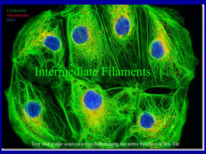
Cytokeratin Mitochondria DNA. Intermediate Filaments. Text and image sources are included using the notes function of this file. The IMF Family. form heterodimers. Assembly. central rod domain. as simple as a bc d efg. bundles of 8 tetramers. Atomic Model of the Dimer.

E N D
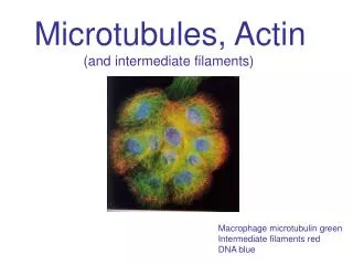
Cytokeratin Mitochondria DNA Intermediate Filaments Text and image sources are included using the notes function of this file
The IMF Family form heterodimers
Assembly central rod domain as simple as abcdefg bundles of 8 tetramers
Cytokeratins in Epithelial Cells Keratins Lamins
Epidermolysis Bullosa Simplex arises from keratin point mutations • Dowling-Meara - widespread blistering • Koebner - blistering confined to hands and feet • Weber-Cockayne - least severe
Normal Skin Squalous cell carcinoma Cancer detection Cytokeratins are normally expresssed in skin but not in the underlying tissues. Antibody staining can reveal hyperplasia and metastasis of epithelial cells into those tissues as a cancer progresses.
NF-H in neurons GFAP in glia Neurofilamentsin Neurons
Active Remodeling (note - short clips are repeated several times in each part of the sequence)
Cytokeratin-GFP dynamics 4 hour sequence
Cytokeratin-GFP dynamics during mitosis 40 minute sequence
Okadaic acid (phosphatase inhibitor) results in cytokeratindisassembly Cytokeratin 13::GFP 200+ minute sequence
Cytokeratin-GFP dynamics require ATP 96 minute sequence
Cytokeratin-GFP dynamics requires microtubules 115 minute sequence
Microinjection of anti-detyrosinated tubulin antibodies collapses the IMF network
Cytokeratin-GFP dynamics doesn’t require micro-filaments 115 minute sequence
Time-lapse Vimentin-GFP Nocodazole collapse Close-up cell edge
Vimentin overlays Distinct from Microfilaments Co-align with Microtubules
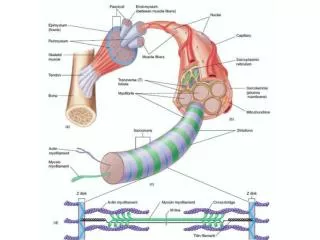
Tensegrity F-actin
Nocodozole-treatment depolymerizes mts and causes lamellipodial retraction