Download
1 / 48
550 likes | 1.34k Views
Elbow( Humeroulnar ) Joint. Presentation by Lindsey Bidleman and Linda McConnell. Components of the Elbow Joint Include…. Surface Anatomy Bones Articular Capsule Cartilage Bursae Ligaments Muscles Nerves Arteries Veins. Surface Anatomy of the Elbow. Cubital Fossa

E N D
Elbow(Humeroulnar) Joint Presentation by Lindsey Bidleman and Linda McConnell
Components of the Elbow Joint Include… • Surface Anatomy • Bones • Articular Capsule • Cartilage • Bursae • Ligaments • Muscles • Nerves • Arteries • Veins Linda
Surface Anatomy of the Elbow • Cubital Fossa • Medial Bicipital Groove • Biceps Tendon • Triceps Tendon • Olecranon • Lateral Epicondyle • Medial Epicondyle • Radial Styloid Process • Ulnar Styloid Process Linda
Surface Anatomy of the Elbow Joint Medial Bicipital Groove Biceps Tendon Cubital Fossa Lateral Epicondyle Medial Epicondyle Olecranon Triceps Tendon Linda
Surface Anatomy of the Elbow Joint Radial Styloid Process Ulnar Styloid Process Linda
Carrying angle Surface Anatomy of the Elbow Joint • When arms are at your sides, palms facing forward, you hands and forearms should be about 5-15 degrees away from your body. • This angle allows your forearms to clear you hips when swinging your arms while walking. Also very important when carrying various objects. • The angle is more pronounced in women than men. Linda
Surface Anatomy of the Elbow JointCarrying Angle: Male vs Female Linda
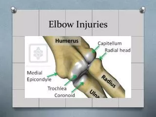
Bones of the Elbow Joint Include… • Humerus- Largest bone in the upper extremity. Articulates with the radius and ulna. • Ulna- The stabilizing bone of the forearm. The medial and longer bone of the two forearms. (Pinky side) • Radius- The lateral and shorter of the two forearm bones. (Thumb side) Linda
Trochlear Notch Ulna Olecranon Process Coronoid Process Radial Notch • Olecranon Process- Big bony projection on proximal end. • Coronoid Process-Prominant elevation on anterior surface. • Trochlear Notch- Articulates with the trochlea of the humerus. • Ulnar Tuberosity- Inferior to the coronoid process. • Radial Notch- Smooth, rounded curve that articulates with the head of the radius. Ulnar Tuberosity Linda Anterior Right Posterior Right
Posterior Right Anterior Right Humerus • Capitulum- Articulates with the head of the radius. • Olecranon Fossa- Big depression on the posterior side of the humerus. • Medial Epicondyle- More prominent than the lateral epicondyle. • Trochlea- Articulates with the trochlear notch of the ulna. • Coronoid Fossa- Superior to the trochlea, the smaller depression in the anterior side of the humerus. • Lateral Epicondyle- Smaller than the medial epicondyle. Coronoid Fossa Medial Epicondyle Lateral Epicondyle Capitulum Olecranon Fossa Trochlea Linda
Radius Head Neck Radial Tuberosity • Head- Smooth, flat surface for articulation with the capitulum of the humerus. • Neck- Narrow part between the head and the radial tuberosity. • Radial Tuberosity- Directly under the head and neck, flat surface. The attachment for the biceps muscle. Linda Anterior Right
Articular Capsule • Articular Capsule is “sleeve like” and surrounds a synovial joint, encloses the synovial cavity, and unites articulating bone. Composed of two layers… • Fibrous Membrane- usually consisting of dense irregular connective tissue that attaches to the periosteum of the articulating bones. • Synovial Membrane- Composed of areolar connective tissue with elastic fibers. Linda
Cartilage • Cartilage is a solid, stretchable type of connective tissue that forms parts of the skeleton where more flexibility and protection are necessary. • Articular Cartilage provides a smooth, low friction gliding surface for free movement for the humerus, radius, and ulna. • Its shiny surface also makes it kind of pretty! Linda
Bursae • Bursae are closed sacs containing fluid , they prevent friction and enable structures to move freely over one another. • Intratendinous Olecranon Bursa- Sometimes present in the tendon of the triceps • Subtendinous Olecranon Bursa- Located between the olecranon and the triceps tendon, just proximal to its attachment to the olecranon • Subcutaneous Olecranon Bursa- Located in the subcutaneous connective tissue over the olecranon Linda
Clinical Awareness of Bursae • Injury can happen to the subcutaneous olecranon bursa by falls on the elbow, and from infraction from abrasions of the skin covering the olecranon, causing the bursa to become inflamed. • Repeated excessive pressure and friction produce a friction called Subcutaneous olecranon bursitis. • Pain is severe during flexion of the forearm • It is easy to treat if the patient follows the “P.R.I.C.E”. Protection, Rest, Ice, Compression, Elevation. Linda
Ligaments: Connect bone to bone • Collateral ligaments of the elbow joint are strong triangular bands that are medial and lateral thickenings of the fibrous layer of the joint capsule • Ulnar Collateral Ligament- Medial and triangular ligament that extends from the medial epicondyle of the humerus to the coronoid process and olecranon of the ulna consisting of three bands… 1. Anterior cord-like band is the strongest 2. Posterior fan-like band is the weakest 3. Slender oblique band that deepens the socket for the trochlea of the humerus Linda
Ligaments Cont… Radial Annular Ligament • Radial Annular Ligament- This ligament encircles and holds the head of the radius in the radial notch of the ulna, and permits pronation and supination of the forearm • Radial Collateral Ligament- Lateral fan-like ligament that extends from the lateral epicondyle of the humerus to the annular ligament of the radius and the radial notch of the ulna. Radial Collateral Ligament Linda
Ligaments Continued Interosseous Membrane- Fibrous connective tissue that joins the shafts of the radius and ulna. Interosseous Membrane Linda
Tis’ the Season http://consumingcostarica.files.wordpress.com/2011/11/turkey.jpg http://www.ebaumsworld.com/pictures/view/82927666/ http://www.andpop.com/2012/10/02/10-funny-turkey-day-photos/ http://sharelike.me/time-and-events/funny-thanksgiving-turkey-cartoon/
Actions of Thanksgiving http://www.google.com/search?q=thanksgiving+praying&client=safari&rls=en&source=lnms&tbm=isch&sa=X&ei=kBWJUpvfNoPj2AXRwYFg&ved=0CAkQ_AUoAQ&biw=980&bih=648#facrc=_&imgdii=_&imgrc=jGWndG_tXGgZlM%3A%3BDXJM2b8TbPyTRM%3Bhttp%253A%252F%252Fcache2.asset-cache.net%252Fxc%252F87834607-family-praying-at-thanksgiving-table-photos-com.jpg%253Fv%253D1%2526c%253DIWSAsset%2526k%253D2%2526d%253D910C62E22B9F47AAC26E85847CFC85B782FF195C3D97418B98B265C480BC42FEE30A760B0D811297%3Bhttp%253A%252F%252Fwww.photos.com%252Froyalty-free-images%252Ffamily-praying-at-thanksgiving-table%252F87834607%3B506%3B336
Elbow Flexion – Forearm Pronation http://admissions.vanderbilt.edu/insidedores/2011/11/thanksgiving-break-so-far/img_0358/
Nerve Supply to the Elbow Brachial Plexus RootsRandy TrunksTravis DivisionsDrinks ChordsCold BranchesBeer B. Branches of Brachial Plexus (Lateral to Medial) MusculocutaneousMoms AxillaryAre RadialReally MedianMad UlnarUsually
Innervations of Elbow • The Musculocutaneous Nerve • Supplies the elbow flexors EXCEPT the brachioradialis • The Radial Nerve • Supplies the elbow extensors • The Median Nerve • Supplies all the pronators of the forearm • The Ulnar Nerve • Runs posterior to the medial epicondyle
The Ulnar Nerve • Known as the “Funny Bone” • Largest nerve that is unprotected by deep tissues, ligaments, muscles, or bones. • The severity of the numbness or pain varies from person to person • Can cause spontaneous paralysis of pinky and lateral ½ of ring finger. • http://youtu.be/ZEcNgyIOO_E
Arteries of the Elbow • Brachial • Anterior interosseous • Ulnar • Superficial palmar arch • Radial • Recurent interosseous • Posterior interosseous
Upper Extremity vascular site significance http://www.nowpublic.com/culture/pulse-rate http://www.papercards.com/sp/CD4934.asp
Location of Brachial Artery • To control hemorrhage • Site where cuff compresses artery against humerus to obtain blood pressure
Veins of the Elbow • Cepthalic • Basilic • Brachial • Median antebrachial • Median cubital • Dorsal venous arch
Venipuncture Upper extremity veins provide best source to obtain blood • It is readily assessable • Veins can be visualized • Quickly cleaned • Does not impede with life activities http://www.oneplaceforspecialneeds.com/main/library_blood_test.html
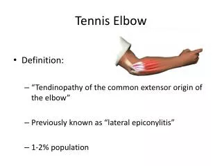
Tennis Elbow • Elbow tendinitis(tennis elbow) is inflammation of the lateral epicondyle. • Occurs most commonly in the extensor carpi radialis brevis, where there is an increase in pain receptors in the area making the region very tender! • Causes of tennis elbow… • The most common cause is the overuse or repetitive strain caused by repeated extension of the wrist against resistance. • Gripping heavy objects • Tennis is also a cause, although the above causes are more common. Linda
Treatment for Tennis Elbow • Goals of treatment • Identify the cause of injury • Reduce pain and inflammation • Gradually return the patient to activity • Treatment • It may take several different types of exercise to completely relieve pain caused by tennis elbow… • Icing to reduce inflammation and pain. • Plenty of rest, but also with a few low grade exercises such as… • Stretching Exercises • Strengthening Exercises • The Real Life Dangers of Tennis Elbow - YouTube Linda
References • You Tube Zach Thurow( April 4th, 2012) Retrieved on November 15th, 2013. The Real Life Dangers of Tennis Elbow - YouTube • Sportsinjuryclinic.net (2013). Tennis Elbow/ Lateral Epicondylitis. Retrieved November 15th, 2013 from http://www.sportsinjuryclinic.net/sport-injuries/elbow-pain/tennis-elbow • A.D.A.M. quality (1997-2013). Carrying Angle of the Elbow- excessive. Retrieved November 15th, 2013. Carrying angle of the elbow - excessive: MedlinePlus Medical Encyclopedia • Wikimedia Commons (April 23, 2013). File: Slide2xzxzxz.JPG. Retrieved on November 16th, 2013. File:Slide2xzxzxz.JPG - Wikipedia, the free encyclopedia • Tortora, G. & Derrickson, B. (2012). Principles of Anatomy & Physiology. (13th ed.). Hoboken, NJ: John Wiley & Sons, Inc. Retrieved November 2013.
References • Moore, L., Agur, A., & Dalley, A. (2011). Essential Clinical Anatomy (4th ed.). Baltimore, MD: Lippincott Williams & Wilkins. Retrieved November 2013. Figures: SA6.3, SA6.4, 6.55, 6.56, 6.57, B6.21 • Clemente, C. (2011). Anatomy A Regional Atlas of The Human Body (6th ed.). Baltimore, MD: Lippincott Williams & Wilkins. Retrieved November 2013. Figures: 88-1l, 88-3l Linda
Quiz on Thursday 1.Biceps Brachii 2.Brachialis 3.Supinator 4.Brachioradialis 5.Pronator Quadratus