Download
1 / 32
340 likes | 1.22k Views
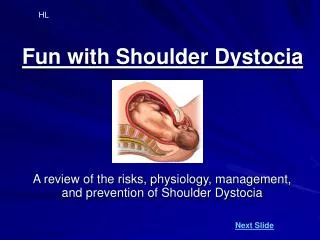
Dystocia & Obstructive Labor. by, Fahad Mohsen Al- Otaibi Supervissor , Ass. Prof. Mohammad Al- Khatim. Objectives. CPD(disproportions) Definitions Etiology Management Soft tissues obstruction Etiology Management Complications Ruptured uterus Etiology Management. Dystocia

E N D
Dystocia& Obstructive Labor by, FahadMohsen Al-Otaibi Supervissor, Ass. Prof. Mohammad Al-Khatim
Objectives • CPD(disproportions) • Definitions • Etiology • Management • Soft tissues obstruction • Etiology • Management • Complications • Ruptured uterus • Etiology • Management • Dystocia • Definition • Etiology • Maternal & fetal complications • Management
Dystocia - Definition Means difficult labor and is characterized by abnormally slow or no progress of labor.
Persistent Occipitotransverse Position • When the head fails to flex and rotate → persists in an occipitotransverse position . • causes : Cephalopelvic disproportion , Altered pelvic architecture with a platypelloid or android pelvis & Relaxed pelvic floor, brought about by epidural anesthesia or multiparty . • management : • If fetal head is at or above the level of the ischial spines or the midpelvis is compromised, when the pelvis is of a platypelloid or android type → Cesarean delivery . • normal size pelvis, uterine contractions are inadequate &the fetus is not macrosomic→ Oxytocic stimulation of labor or by Manual rotation using the fingers of the examiner's hand or forceps rotation using Kielland forceps .
Manual rotation using the fingers of the examiner's hand or forceps rotation using Kielland forceps
Macrosomia • Definition : fetal weight ≥ 4.0 or 4.5 kg . • Risk factors : more in male fetus , previous macrosomic infant , excessive maternal weight gain , multiparty ,postterm pregnancies , GDM . • Diagnosis : clinically & US . • Complications : prolonged pregnancy , cephalopelvic disproportion , obstructed labour , shoulder dystocia , birth injuries &future baby obesity . • Management • Proper prenatal care to prevent macrosomia and diagnose it before labor . • Caesarean delivery .
an enlargement of the fetal head due to accumulation of excessive cerebro-spinal fluid (C.S.F) within the ventricles. • 0.5-1.8 per 1000 births . • Causes : Genetic aberration as trisomies , infections: as cytomegalovirus, toxoplasmosis and rubella or idiopathic . • Diagnosis : US , large head with biparietal diameter >12 cm , dilated cerebral lateral ventricles , small face in relation to the head size & the thickness of cerebral cortex . • Complications : obstructed labor with its sequel as rupture uterus & fetus: neurological manifestations and low growth rate. Hydrocephalus
Management Hydrocephalus • intrauterine ventriculo-amniotic shunt: to drain the CSF from the cerebral ventricles into the amniotic cavity . • In Cephalic presentation , if the cervix is dilated→ transcervical aspiration by a needle or perforation through a gaping suture or fontanelle is done. • if the cervix is not dilated→ transabdominal aspiration with the aid of ultrasonic visualization , traction on the collapsed head can then applied forceps. • In Breech presentation: • CSF is drainedthrough: Spinal tapping through spina bifida if present. • cesarean section to avoid the risk of infection, which can result from transvaginal or trans-abdominal drainage
… Management Hydrocephalus • ascitesin the fetal abdomen or enlargement of fetal organs, such as the bladder or liver, may result in unexpected dystocia after the fetal head is delivered →Ascitic fluid or urine from a bladder removed by transabdominal drainage with a needle before vaginal delivery . • Cesarean delivery may be indicated if the fetal abdomen cannot be sufficiently decompressed. • Postpartum:the living newborn should be referred for shunt operation.
Cephalopelvic Disproportion • Definition: Maternal bony pelvis is not of sufficient size and of appropriate shape to allow the passage of the fetal head. • Maternal causes : Contractions of one of the planes of the pelvis . • Fetal causes :Excessively large fetal head or abdomen & Abnormal positioned fetus . • Degrees & management
Contracted pelvis Definition: Anatomical definition: It is a pelvis in which one or more of its diameters are reduced below the normal by one or more centimeters. Obstetric definition: It is a pelvis in which one or more of its diameters are reduced so that it interferes with the normal mechanism of labor.
Diagnosis Contracted pelvis
Management Contracted pelvis
Contracted pelvis Complications Maternal Fetal • During labour: • Slow cervical dilatation and prolonged labour. • Premature rupture of membranes and cord prolapse. • Obstructed labour and rupture uterus. • Injury to pelvic joints or nerves from difficult forceps delivery. • Postpartum hemorrhage. • During pregnancy: • ↑retroverted gravid uterus. • Malpresentations. • Pendulous abdomen • Nonengagement. • Pyelonephritis due to more compression of the ureter. • Intracranial hemorrhage. • Asphyxia. • Fracture skull. • Nerve injuries. • Intra-amniotic • infection
Obstructed labor • It is the arrest of vaginal delivery of the fetus due to mechanical obstruction . • Maternal causes : • Bony obstruction: • Contracted pelvis. • Tumors of pelvic bones. • Soft tissue obstruction: • Uterus: impacted subserouspedunculated fibroid, constriction ring opposite the neck of the fetus. • Cervix: cervical dystocia. • Vagina: septa, stenosis, tumors. • Ovaries: Impacted ovarian tumors.
… Obstructed labor • Fetal causes : • Malpresentations and malpositions: • Persistent occipito transverse position and arrest, • Brow, Shoulder, Impacted breech • Large sized fetus (macrosomia). • Congenital anomalies: • Hydrocephalus. • Fetal ascitis. • Fetal tumors. • Twins.
Obstructed labor Diagnosis 1-history: • prolonged labor, • frequent and strong uterine contractions, • Rupture membranes. 2-Examination General examination: • It shows signs of maternal distress as: exhaustion, high temperature (³ 38oC) & rapid pulse . • Signs of dehydration: dry tongue and cracked lips.
Obstructed labor … Diagnosis Abdominal examination: • The uterus: hard ,tender & frequent strong uterine contractions with no relaxation. • The fetus: fetal parts cannot be felt easily & FHS are absent or show fetal distress due to interference with the utero-placental blood flow. Vaginal examination: • Cervix: is fully or partially dilated. • The membranes: are ruptured. • The presenting part: is high and not engaged or impacted in the pelvis.
Obstructed labor Diagnostic criteria • Failure of the presenting part to descend • Partogram will show above 2 parameters
Obstructed labor Complication • 1-Maternal: • Maternal distress • Rupture uterus. • Necrotic vesico-vaginal fistula. • Infections as chorioamnionitis and puerperal sepsis. • Postpartum hemorrhage due to injuries or uterine atony. • 2-Fetal: • Asphyxia. • Intracranial hemorrhage. • Birth injuries. • Infections.
Obstructed labor Management • Preventive measures: Careful observation, proper assessment, early detection and management of the causes of obstruction. • Prophylactic measure: 1-Antibiotic(At risk of tetanus, Clostridium Welchii) 2-ttt of shock(Dehydration, hypovolemic, septic) • Curative measures: Caesarean section is the safest method
Uterine Dehiscence or Rupture Definition: Dehiscence:is define as: separation of a lower uterine scar that does not penetrate the serosa and rarely causes significant hemorrhage. Rupture is defined as complete separation of the uterine wall and may lead to significant hemorrhage and fetal distress. • Incidence • Uterine rupture occurs in 0.2% to 1% of patients with a previous low-segment transverse cesarean section • 4% to 9% of patients with a prior uterine active segment incision (classical or T-incision) • One-third of women with a history of previous classical cesarean section who experience rupture do so before onset of labor
Uterine Dehiscence or Rupture Risk factors • previous uterine surgery • cesarean section, myoectomy, previous resection of a cornual ectopic pregnancy, or previous uterine perforation • Induction of labor with prostaglandin agents in the setting of a history of a previous cesarean further increases the risk of rupture • Other risk factors are internal version or extraction, operative delivery, and trauma
Uterine Dehiscence or Rupture Diagnosis • Fetal bradycardia is clinically manifested in 33% to 70% of cases • Fetal distress may be initial presentation in catastrophic uterine rupture • In milder cases the initial presentation is a simple rise in fetal station or change in position for fetal heart monitor placement. • fetal parts may be more easily palpable abdominally • Maternal signs and symptoms include constant abdominal pain , shock ( hypotension and tachycardia), cessation of uterine contractions, uterine tenderness, or a change in uterine shape, vaginal bleeding
Uterine Dehiscence or Rupture Management • Emergent laparotomywith delivary of the infant and repair of the uterine rupture if small thin edge no bleeding not extended to uterine artery • If not hysterectomy .
Summary • Dystocia means difficult labor that caused by many conditions e.g. macrosomia & hydrocephalous . • many pelvic problems can lead to persistent occipitotransverse position that can be managed by C-S or stimulation of labor . • macrosomia > 4kg with high relation to GDM . • hydrocephalous can lead to obstructive labor & fetal low growth rate . • Cephalopelvic disproportion is maneged according to its degree either by vaginal delivery or C-S . • contracted pelvis causes either in pelvis or in spine . • Obstructed labor caused by bon,soft tissue obstruction or malpresentations , macrosomia & congenital anomalies . • C-S is the safest method in management of obstructed labor . • Uterine dehiscence (separation of a lower uterine scar without hemorrhage ) but rupture (complete separation of the uterine wall with significant hemorrhage and fetal distress ). • managed by either emergent laparotomy or hysterectomy .
Thank You Any Question