Download

1 / 26
290 likes | 511 Views
IHE Mammography: Workflow, Display, and Dose Monitoring. Christoph Dickmann, Siemens Healthcare RSNA 2008 Informatics Courses. FFDM - not just another modality. Full Field Digital Mammography (FFDM) imaging has specific requirements: High patient throughput (e.g. 300 patients/day)

E N D
IHE Mammography: Workflow, Display, and Dose Monitoring Christoph Dickmann, Siemens Healthcare RSNA 2008 Informatics Courses
FFDM - not just another modality Full Field Digital Mammography (FFDM) imaging has specific requirements: • High patient throughput (e.g. 300 patients/day) • “On-line” work-up, screening/ diagnostic • Common use of CAD (Computer Aided Detection) • Double (blind) reading • Importance of prior study comparisons • Image size, orientation, layout
Integration issues to be solved • Variances among vendors’ image data • Two types of image data • Hanging may look different on workstations • CAD markers may look different on workstations • Add supplemental images • Change procedure information at Modality • Hide low-quality or incorrectly labeled images from further use
Mammography Environment Workstation Mammo IS FFDM Modality Printer CAD “For Processing” Images Archive “For Presentation” Images Mammo CAD Report Procedure, Worklist, Procedure Step Patient dose information
Mammography Image ProfileValue proposition • Systems support DICOM objects • enhanced for interoperability/ quality aspects • FFDM modalities provide adequate information to facilitate downstream applications • Image display and printing operations enable safe, effective and efficient diagnoses • View and print images from multiple FFDM vendors consistently
Acquisition ModalityRich image information for high-quality display • Send both - FOR PROCESSING and - FOR PRESENTATION images with reference that ties them together • Additionally required DICOM attributes, e.g. • Acquisition date & time, operator’s name • Detector ID, software version • Exposure time, filter material, positioner angle • Detection and indication of tissue vs. air • Partial View Option - breast is larger than detector • Similar for film digitizers (less requirements)
Image DisplayConsistent presentation & screen layout R CC L CC R MLO L MLO Calibration • DICOM Grayscale Standard Function Image hanging determined by: • View Type (e.g. CC vs. MLO) • View Type modifiers (e.g. Spot, Mag) • Laterality • Patient Orientation IHE does not use for hanging • Series or study descriptions • Image order as acquired • Image orientation as acquired
Image DisplayConsistent pixel sizes (un)magnified 4096 Acquisition Matrix: Image Receptor e.g. 20,000,000 Pixels Display Matrix: Monitor e.g. 5,000,000 Pixels 5024 2560 2048
Image DisplayConsistent sizing also with different detectors R L R L R L L R Prior exam from vendor A’s system Current exam from vendor B’s system Different resolution Different pixel matrix Fit images to same size: • Relative (e.g. fit to viewport) • Physical (true size) • 1:1 pixels (detector : screen)
Image DisplayContrast, annotations, CAD marks Contrast Ability to use linear and sigmoid LUT Tissue vs. air detection: Window/ Center adjustments are not applied to air Image annotations • Patient identification • Equipment identification • Technical factors • View: laterality, view, view modifier CAD marks from Mammography CAD SR • One or more sets of CAD marks • Fit to image orientation
Printing: Composer, Server Quality, annotations Minimal borders at chest wall • True Size printing • Physical object size • Composer sends Requested Image Size • Burn in a ruler • Justify chestwall • print with 5mm borders or less • Use display annotations • Use Presentation LUT • Use maximum density values • Support 12 bit pixel depth
Mammo Acquisition Workflow Value proposition Now that I can create and consistently display Mammography images, how can image acquisition be handled reliably for routine and “exceptions”? • Routine: same as Radiology Scheduled Workflow • Clearly record what is performed (differently) at the Modality coded values • Correctly display images from acquisitions of separate procedure steps • Mark images of insufficient quality • Correct certain image labeling
Mammography EnvironmentAcquisition Workflow view Mammo CAD Report “For Presentation” Images Procedure, Worklist, Procedure Step Mammo IS Workstation FFDM Modality Proc. Step Worklist,Proc. Step Procedure Archive
Mammo Acquisition Workflow Actors & Transactions – the IHE environment DSS = Department System Scheduler. Abbreviations on subsequent slides: OF = Order Filler. (M)PPS = (Modality) Performed Procedure Step
Mammo Acquisition WorkflowSupplemental views to a completed exam MPPS forwarded MPPS in progress MPPS completed Query/ Retrieve screening and implant images Store supplemental images Archive Modality DSS/ OF * Workstation Scenario: Supplemental image of implant displacement after screening • Append case: Add image to current Procedure, e.g. “Screening bilateral” • Tech selects from configured Modality protocol settings • Modality sends a new MPPS with the protocol codes • DSS/OF may use these protocol codes, e.g. for notifying the Clerk/ Tech • Image Display selects presentation layout based on procedure/ protocol/ reason codes * Note: For simplicity reasons, the diagrams show the PPS Manager as part of the DSS/OF
Mammo Acquisition WorkflowFinish acquisition at another Modality Query MWL MPPS forwarded MPPS in progress MPPS completed Query/ Retrieve images from both Modalitiesas part of the same Study Store images Query MWL (patient-based) MPPS in progress/ completed Store images Modality 1 Archive DSS/ OF Workstation Modality 2 Scenario: Supplemental images acquired later, at another Modality • Avoid unscheduled acquisition with manual reconciliation at DSS/OF • It all depends on DSS/OF worklist behavior • Modality queries Worklist (MWL) based on: Patient name/ ID, Accession #, Procedure ID
Mammo Acquisition WorkflowConvert procedure MPPS in progress MPPSforwarded MPPS completed Query/ Retrieve imagesfor bilateral diagnostic exam Store images of R breast Archive Modality DSS/ OF Workstation Procedure update: unilateral bilateral Scenario: Convert diagnostic procedure from unilateral to bilateral • Radiologist reviews images of left breast while patient still present.Radiologist decides that images of right breast are needed, too. • Append case: Add images to current Procedure that is changed to “Diagnostic Mammography, bilateral” • Tech adds reasons for changing the Procedure (new DICOM attribute: CP 886) • Tech selects from a configurable Modality protocol set • Modality sends a new MPPS with new procedure & protocol codes • DSS/ OF detects procedure change and updates Image Manager/ Archive.
Mammo Acquisition WorkflowRecall – patient returns for subsequent exam Query MWL (on recall) MPPS forwarded MPPS Query/ Retrieve images from original and recall study Store images (recall study) Archive Modality DSS/ OF Workstation Scenario: Screening recall for a unilateral diagnostic mammogram • Radiologist assesses acquired images as “incomplete” (BIRADS 0). Patient has left and needs to come back for additional imaging. • DSS/OF schedules a new Procedure (for the screening Order) with • a specific Reason for Requested Procedure code value • a reference to the original Study (Referenced Study Sequence) • Regular acquisition at Modality • Image Display presents both Studies based on Reason code and Study reference
Mammo Acquisition WorkflowReject images of insufficient quality MPPS forwarded MPPS (Modality) orCreator PPS AvailabilityNotification Q/R A) Rejected images not part of query result Store rejection KOS Archive DSS/ OF Workstation Modality or QA workstation A) Rejected images are hidden B) Rejected images are hidden or displayed B) Rejected images are provided Scenario: Mark images as rejected for quality reasons • After completed acquisition, images are considered of insufficient quality • Modality or Evidence Creator create a Key Object Selection (KOS) object • Title is “Rejected for Quality Reasons” and explained by a Modifier • References to all rejected images • Image Archive is configurable to provide or hide rejected images and KOS • Image Display is configurable to present or hide rejected images and KOS
Mammo Acquisition WorkflowCorrect view labels of images PPS forwarded MPPS (Modality) orCreator PPS Availability Notification Q/R: incorrect imagesare not part of query result Store rejection KOSStore corrected image(s) Archive DSS/ OF Workstation Modality or QA workstation Incorrect images are hidden at Q/R Scenario: View, laterality or protocol are corrected • After completed acquisition, incorrect view labeling is detected • Modality or Evidence Creator create corrected images • Copy most values except e.g. View Codes, View Modifiers, Laterality, Protocol • Correct references from “For Presentation” to “For Processing” images • Evidence Creator records its identifying data (Contributing Equipment Sequence) • Modality or Evidence Creator create a Key Object Selection (KOS) object • Title is “Rejected for Patient Safety Reasons” • References to all incorrect images (For Presentation & For Processing) • Image Archive & Image Display hide KOS and incorrect images
Mammo Acquisition WorkflowSummary • Extend routine Scheduled Workflow • Use of Protocol and Reason codes (DSS/OF, Modality) • Scheduled and Performed Procedure Codes can vary.Never empty Procedure or Protocol Code Sequences. • Modality supports Patient-based Worklist Query • Image Quality Assurance results • “Low quality” images can be marked or hidden • Incorrectly labeled images can be corrected
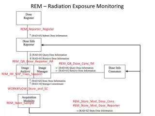
Radiation Dose Monitoring in Mammography NationalRegistry some text: # Numerical Details 12.2 14.5 11.8 7.6 9.5 10.9 some text: # Numerical Details 12.2 14.5 11.8 7.6 9.5 10.9 some text: # Numerical Details 12.2 14.5 11.8 7.6 9.5 10.9 some text: # Numerical Details 12.2 14.5 11.8 7.6 9.5 10.9 some text: # Numerical Details 12.2 14.5 11.8 7.6 9.5 10.9 Numer 12.2 14.5 9.5 10.9 some text: # Numerical Details 12.2 14.5 11.8 7.6 9.5 10.9 some text: # Numerical Details 12.2 14.5 11.8 7.6 9.5 10.9 some text: # Numerical Details 12.2 14.5 11.8 7.6 9.5 10.9 some text: # Numerical Details 12.2 14.5 11.8 7.6 9.5 10.9 some text: # Numerical Details 12.2 14.5 11.8 7.6 9.5 10.9 Outlier: # Performing Phys. Over Target: 12.2% Outlier: # Performing Phys. Over Target: 12.2% some text: # Numerical Details 12.2 14.5 11.8 7.6 9.5 10.9 Outlier: # Performing Phys. Over Target: 12.2% Dose Analysis & Reporting Archive FFDM Modality also implements the Acquisition Modality in the Radiation Exposure Monitoring IHE Profile
Radiation Dose Monitoring in Mammography • FFDM Modality can act as an Acquisition Modality in the IHE Radiation Exposure Monitoring Profile • Modality reports patient dose • Archive/ PACS receive and store dose reports • RIS, Workstations, etc. process or display these reports • Dose Reports are DICOM Dose Reports for projection X-ray • Required: dose of irradiation events • Optional: accumulated dose over Study or Series • Mammography Dose • Organ-specific: laterality, fibroglandular dose, breast composition, compression • Exposure: time, KVP, mA, entrance exposure at reference point
Summary • IHE Mammography Profiles make FFDM Modalities, PACS, Viewing Stations and Printers interoperable: • Consistently use rich data in DICOM Mammography images • Consistent pixel / image sizing • Consistent windowing • Consistent and correct image layout on screen or film • Exactly defined procedures and workflow steps • Flexibility in adding / changing workflow steps • IHE Radiation Exposure Profile enables monitoring radiation doses in Mammography
Take home • Mammography User Handbookwww.ihe.net/Resources/handbook.cfm • Understand your interoperability issues • Help for making purchase decisions • Technical Frameworkswww.ihe.net/Technical_Framework/ • Technical requirements • SBI 2009 educational events