Download
1 / 42
450 likes | 748 Views
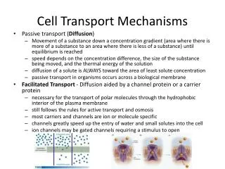
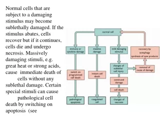
Mechanisms of Cell Death. Etiology of cell death. Major Factors Accidental Genetic Necrosis Apoptosis. Necrosis: The sum of the morphologic changes that follow cell death in a living tissue or organ Apoptosis:

E N D
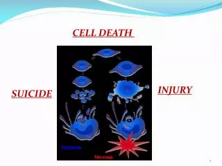
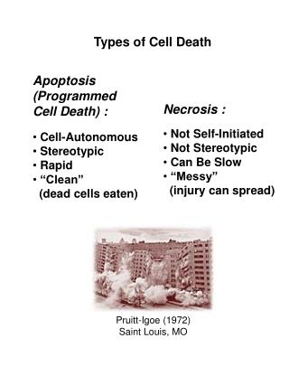
Etiology of cell death Major Factors Accidental Genetic Necrosis Apoptosis Necrosis: The sum of the morphologic changes that follow cell death in a living tissue or organ Apoptosis: a physiological process that includes specific suicide signals leading to cell death
Distinct modalities of cell death Cell Death Differ. 2009, 16(1): 3–11
The road to necrosis Homeostatic ‘steady state’ Cellular adaptations Reversible cell injury Irreversible cell injury Cell death Necrosis
Apoptosis: a physiological response to specific suicide signals, or lack of survival signals Necrosis: a pathological response to cellular injury www.chembio.uoguelph.ca
APOPTOSIS AS A PHYSIOLOGICALLY IMPORTANT PROCESS In addition, there are often apoptotic centers in tumors, accounting for the paradox of slow gross enlargement in the face of rapid cell proliferation, and the rare spontaneous remission. www.chembio.uoguelph.ca
APOPTOSIS in C.elegans C.elegans genome: 19099 genes (790 seven-pass transmembrane receptors, 480 zinc finger proteins, and 410 protein kinases) The life cycle of C. elegans from egg to sexual maturity (and new eggs) is about 3 days ced-1, -3, -4, and -9 (Cell death determining) proteins in C.elegans are closely related to mammalian apoptosis-regulating genes The adult hermaphrodite consists of exactly 959 somatic cells of precisely determined lineage and function. Individual cells are named and their relationships to their neighbors are known Overall, the 959 somatic cells of adult C.elegans arise from 1090 original cells; exactly 131 somatic cells undergo programmed cell death in the wild type worm Of the 1090 cells, 302 are neurons, and many of the programmed deaths also lie in the neuronal lineage www.chembio.uoguelph.ca
Autophagic cell death (type II programmed cell death) – meaning that the cytoplasm is actively destroyed long before nuclear changes become apparent; Classical apoptotic cell death – meaning that the chromatin marginates and the cell and nucleus fragment before morphological changes are seen in intracellular organelles Nature Immunology4, 416 - 423 (2003)
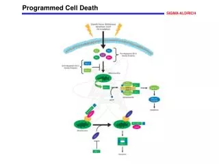
APOPTOSIS SIGNALS Mitochondria- dependent apoptosis Death Receptor- dependent apoptosis Caspase- dependent apoptosis Caspase- dependent apoptosis Caspase- independent apoptosis Caspase- independent necrosis Nature Reviews Cancer2; 647-656 (2002)
Galluzzi et al. Cell Death & Differentiation (2012) 19, 107–120
In response to multiple intracellular stress conditions (e.g., DNA damage, cytosolic Ca2þ overload), pro-survival and pro-death signals are generated and converge to a mitochondrion-centered control mechanism. When lethal signals prevail, mitochondrial outer membrane permeabilization (MOMP) occurs and leads to mitochondrial trans-membrane potential (Dcm) dissipation, arrest of mitochondrial ATP synthesis and Dcm-dependent transport activities. Moreover, the respiratory chains gets uncoupled, leading to generation of reactive oxygen species (ROS), and proteins that are normally confined within the mitochondrial inter-membrane space (IMS) are released into the cytosol. Among these, cytochrome c (CYTC) drives – together with the cytoplasmic adaptor protein APAF1 and dATP – the assembly of the so-called apoptosome, a multi-protein complex that triggers the caspase-9-caspase-3 proteolytic cascade. Direct IAP-binding protein with low pI (DIABLO, also known as second mitochondria-derived activator of caspases, SMAC) and high temperature requirement protein A2 (HTRA2) facilitate caspase activation by sequestering and/or degrading several members of the inhibitor of apoptosis protein (IAP) family. On the contrary, apoptosis-inducing factor (AIF) and endonuclease G (ENDOG) function in a caspase-independent manner by relocating to the nucleus and mediating large-scale DNA fragmentation. Of note, the serine protease HTRA2 also contributes to caspase-independent apoptosis by cleaving a wide array of cellular substrates (including cytoskeletal proteins). IM, mitochondrial inner membrane; OM, mitochondrial outer membrane; PTPC, permeability transition pore complex Caspase-dependent and -independent ‘intrinsic apoptosis’ Galluzzi et al. Cell Death & Differentiation (2012) 19, 107–120
Extrinsic Apoptosis: apoptotic cell death that is induced by extracellular stress signals that are sensed and propagated by specific transmembrane receptors Upon FAS ligand (FASL) binding, the cytoplasmic tails of FAS (also known as CD95, a prototypic death receptor) trimers recruit (among other proteins) FAS-associated protein with a death domain (FADD), cellular inhibitor of apoptosis proteins (cIAPs), c-FLIPs and pro-caspase-8 (or -10). This supramolecular platform, which has been dubbed ‘death-inducing signaling complex’ (DISC), controls the activation of caspase-8 (-10). Within the DISC, c-FLIPs and cIAPs exert pro-survival functions. However, when lethal signals prevail, caspase-8 gets activated and can directly trigger the caspase cascade by mediating the proteolytic maturation of caspase-3 (in type I cells) or stimulate mitochondrial outer membrane permeabilization (MOMP) by cleaving the BH3-only protein BID (in type II cells). Extrinsic apoptosis can also be ignited by dependence receptors like DCC or UNC5B, which relay lethal signals in the absence of their ligand (netrin-1). In the case of DCC and UNC5B, the pro-apoptotic signaling proceeds through the assembly of a DRAL- and TUCAN- (or NLRP1-) containing caspase-9-activating platform or by the dephosphorylation-mediated activation of death-associated protein kinase 1 (DAPK1) by UNC5B-bound protein phosphatase 2A (PP2A), respectively. DAPK1 can mediate the direct activation of executioner caspases or favor MOMP. tBID, truncated BID Galluzzi et al. Cell Death & Differentiation (2012) 19, 107–120
Regulated Necrosis: necrosis can occur in a regulated manner in addition to spontaneous cell death Upon tumor necrosis factor a (TNFa) binding, the cytoplasmic tails of TNF receptor 1 (TNFR1, a prototypic death receptor) trimers recruit TNFR-associated death domain (TRADD), receptor-interacting protein kinase 1 (RIP1), cellular inhibitor of apoptosis 1 (cIAP1), cIAP2, TNFR-associated factor 2 (TRAF2) and TRAF5. Within the so-called complex I, RIP1 is polyubiquitinated by cIAPs, thereby providing a docking site for the recruitment of transforming growth factor b (TGFb)-activated kinase 1 (TAK1), TAK1-binding protein 2 (TAB2) and TAB3 (which together deliver a pro-survival signal by activating the transcription factor NF-kB). In some patho-physiological and experimental settings, and in particular when caspase-8 is absent or when caspases are inhibited by pharmacological agents, cylindromatosis (CYLD)-deubiquitinated RIP1 engage in physical and functional interactions with its homolog RIP3, ultimately activating the execution of necrotic cell death. Regulated necrosis can also be induced by alkylating DNA damage (possibly by the overactivation of poly(ADP-ribose) polymerase 1, PARP1). In some (but not all) instances, regulated necrosis requires the kinase activity of RIP1, that is, it can be blocked by the RIP1-targeting compounds necrostatins. FADD, FAS-associated protein with a death domain Galluzzi et al. Cell Death & Differentiation (2012) 19, 107–120
Mitotic catastrophe: • cell death occurring in mitosis • cases of cell death that are triggered by aberrant mitosis and executed either during mitosis or in the subsequent interphase • might not even constitute a bona fide cell death executioner mechanism, but an onco-suppressive pathway that precedes and is distinct from, yet operates through, cell death or senescence (a) In the absence of chemical and genetic perturbations of the mitotic apparatus (including chromosomes and the molecular machinery that ensures their faithful segregation), cells progress through the different phases of the cell cycle to generate a diploid offspring. On the contrary, if chromosomal defects or problems affecting the mitotic machinery are sensed during the M phase, cells become arrested in mitosis due to the activation of mitotic catastrophe (b–d). These cells can undergo different fates: they can die without exiting mitosis (b), reach the G1 phase of the subsequent cell cycle (through a phenomenon that is known as mitotic slippage) and then die (c), or exit mitosis and undergo senescence (d). Irrespective of this diversity of outcomes, mitotic catastrophe can be defined as an oncosuppressive mechanism that precedes and is distinct from, but operates through, cell death and senescence Galluzzi et al. Cell Death & Differentiation (2012) 19, 107–120
Autophagic cell death: instances of cell death that are accompanied by a massive cytoplasmic vacuolization In response to stress and during development, eukaryotic cells often activate autophagy, a mechanism whereby organelles and portion of the cytoplasm are sequestered in double-membraned vesicles (autophagosomes) that are delivered to lysosomes for degradation. Stress-induced autophagy most often exerts cytoprotective functions and favors the re-establishment of homeostasis and survival (a). In this setting, pharmacological or genetic inhibition of autophagy accelerates cell death. On the contrary, these interventions frequently inhibit developmental cell death, indicating that autophagy also constitutes a lethal mechanism that mediates ‘autophagic cell death’ (b) Galluzzi et al. Cell Death & Differentiation (2012) 19, 107–120
The target sequence for Ced-3 and caspases (Cys catalytic Asp targeting proteases) consists of a tetrapeptide with C-terminal Asp (D).
Caspase cascades and their inhibitors • Formation of multicomponent complexes triggers initiator caspase dimerization • sufficient for their activation: • DISC: Death-inducing signaling complex • Apoptosome • (PIDD)osome: p53-induced protein with a death domain Cell Death and Differentiation (2011) 18, 1441–1449
In vivo substrates of effector caspases www.chembio.uoguelph.ca
Mitochondria play a central role in mediating the apoptotic signal Mitochondria-free cytoplasm would not induce apoptosis in vitro Cytochrome c-neutralizing antibodies block apoptosis Cytochrome c is an abundant protein of the mitochondrial inner membrane, and acts as an electron transport intermediate. a and b type cytochromes are inaccessible components of large complexes, but cytochrome c is monomeric, freely diffusible in the inner membrane, and in equilibrium between inner membrane, inter-membrane space and cristae. The events of apoptotic activation lead to alterations in permeability of the mitochondrial membrane pore proteins and release of cytochrome c. Initial release of cytochrome c occurs by a highly specific process, involving proteins of the Bcl-2 family www.chembio.uoguelph.ca
Signaling leading to activation of mitochondria-related apoptosis Death receptors of the TNFR family, as well as various oxidants, detergents and chemotherapeutic drugs, induce the release of active cathepsins from the lysosomal compartment. These cathepsins cleave Bid, which can then mediate cathepsin-induced MPT. Disruption of the cytoskeleton leads to the release of the BH3 domain–only proteins Bim and Bmf. DNA damage induced by radiation or various chemotherapeutic drugs induces the p53-mediated transcription of genes encoding Bax, BH3 domain–only proteins (Noxa or Puma), proteins involved in ROS generation and cathepsin D. ER stress results in the release of calcium, which may cause direct mitochondrial damage or activate Bax through calpain-mediated cleavage. Various death stimuli, mediated through death receptors, trigger the production of lipid second messengers (such as ganglioside (GD3), arachidonic acid (AA) and ceramide) that are involved in MPT and mitochondrial damage. Depending on the stimulus and the type of cell, as well as the metabolic status of the cell, MPT leads to either caspase-mediated apoptosis or caspase-independent PCD. Nature Immunology4, 416 - 423 (2003)
Bcl-2 family: Pro-Life and Pro-Death factions Bcl-2 and its closest relatives Bcl-XL, Bcl-w and Ced-9 are a-helical proteins having all four BH domains and are pro-survival. They suppress cytochrome c release, and are oncogenic when overexpressed. However, Bcl-XS, a splice variant of Bcl-XL having BH4 but lacking BH1 and BH2 is pro-apoptotic. Bax and Bak lack the BH4 domain, and are pro-apoptotic. Bax expression is stimulated by p53, a mechanism for pro-apoptotic action of p53. Ectopic or overexpression of Bax induces cytochrome c release and apoptosis, and addition of Bax to mitochondria in vitro induces cytochrome c release. The BH3-only sub group are strongly pro-apoptotic, and include Bim, Bik and Egl-1, which only have the 18-residue BH3 and the transmembrane region, while Bad and Bid only have BH3. The helical BH3 element allows for homo- and heterodimerization between family members. The non-homologous regions of BH3-only proteins could provide links to apoptotic signaling systems. www.chembio.uoguelph.ca
Bcl-2 family: Pro-Life and Pro-Death factions Effector caspases Caspase-9 www.chembio.uoguelph.ca
Mechanisms of mitochondrial outer membrane permeabilization during cell death. AIF: apoptosis inducing factor; ANT: adenine nucleotide translocase; CL: cardiolipin; Cyt c: cytochrome c; CyD: cyclophilin D; CsA: cyclosporin A; IMM: inner mitochondrial membrane; MPT: mitochondrial permeability transition; OMM: outer mitochondrial membrane; VDAC: voltage-dependent anion channel. Orrenius et al., Ann Rev Pharmacol Toxicol 2007
Mitochondria permeability transition can trigger caspase-dependent and caspase-independent programmed cell death (PCD): • Mitochondrial damage leads to the release of numerous mitochondrial proteins that mediate PCD. • Release of cytochrome c triggers caspase activation and classic apoptosis. • Smac (also known as Diablo) and Omi assist cytochrome c–induced caspase activation by counteracting caspase inhibitory factors (IAPs). • AIF triggers a caspase-independent death pathway that culminates in DNA fragmentation and chromatin condensation characteristic of apoptosis-like PCD. • EndoG cleaves DNA and induces chromatin condensation • The serine protease activity of Omi can mediate caspase-independent cellular rounding and shrinkage without changes in the nuclear morphology • Calcium and ROS can lead to severe mitochondrial dysfunction and necrosis-like PCD either directly or through autophagy of damaged mitochondria. Autophagy also may be associated with cathepsin activation and so can result in apoptosis-like PCD. Nature Immunology4, 416 - 423 (2003)
Survival mechanisms downstream of cytochrome c 1. Sequestration by heat shock proteins: Apaf1 interacts with heat shock proteins hsp70 and hsp90. Hsp70 directly sequesters CARD, and blocks caspase-9 recruitment, and possibly assembly of the oligomeric apoptosome as well. Hsp90 also associates with the monomeric Apaf1, and may represent a significant fraction of the normal autoinhibited state. Hsp90 appears to compete with cytochrome c for binding, suggesting action at an earlier step than hsp70. 2. Direct inhibition of the caspase catalysis by Inhibitor of Apoptosis Proteins (IAPs): Inhibitor of apoptosis proteins (IAPs) represent the final line of defense against apoptosis, and act by binding directly to the substrate site of caspases Smac/DIABLO: the mitochondrial answer to IAPs: Mitochondria initiate the apoptosis cascade by releasing cytochrome c, but this effect could be nullified if IAP were allowed to maintain their inhibition of caspases. The apoptotic signal is instead sustained by the release of Smac/DIABLO (second mitochon-drial activator of caspase/direct IAP binding protein of low pI), which binds to and antagonizes the IAPs. www.chembio.uoguelph.ca
DEATH RECEPTORS: Pathways linking external signal receptors to caspase-8 A variety of cell surface receptors related to TNF-R (tumor necrosis factor receptor) interact with the apoptotic activation system. The intracellular portion of the receptor carries a specific protein interaction domain called the death domain, DD. The DD is activated by proximity, brought about when bound extracellular ligand induces receptor oligomerization. Activation can also be induced in absence of ligand by artificial cross-linking of the receptor. Clustered receptor DDs recruit a variety of DD-containing adapters, of which FADD, Fas-associated death domain protein (also known as MORT1) bridges to a second protein interaction domain, DED, or death effector domain. The cluster of FADD-DEDs recruits procaspase-8, which also carries DEDs at its N-terminus (corresponding to the CARDs on Procaspase-9). Procaspase-8 is activated to Caspase-8 by proximity-induced self-cleavage. Procaspase-10 is the only other caspase with DED boxes, and may substitute for Caspase-8 in some cases. In some cells, TNF receptors associate with adaptors linked to cell proliferation or inflammatory signaling pathways, and may induce anti-apoptotic c-IAPs. www.chembio.uoguelph.ca
Death receptor–triggered caspase-dependent and caspase-independent pathways The death receptor is stimulated by ligand-induced activation of the receptor trimer. The receptor death domains (DDs) of Fas then recruit FADD and RIP1 to the receptor complex. After recruitment to FADD through interactions between their death effector domains (DEDs), caspase-8 and caspase-10 are activated and trigger effector caspases, either directly or through a Bid-mediated mitochondrial pathway (activation of Apaf-1 and caspase-9). FADD and RIP initiate a caspase-independent necrotic pathway mediated by the formation of, most probably, mitochondrion- or cPLA2-derived ROS. TNFR1 signaling differs from Fas signaling in the following steps: first, binding of FADD and RIP to the receptor complex requires the adaptor protein TRADD; and second, the RIP1-mediated necrotic pathway is inhibited by FADD and activated caspase-8 Nature Immunology4, 416 - 423 (2003)
“Gentamicin is an aminoglycoside antibiotic widely used against infections by Gram-negative microorganisms. Nephrotoxicity is the main limitation to its therapeutic efficacy. Gentamicin nephrotoxicity occurs in 10–20% of therapeutic regimes. A central aspect of gentamicin nephrotoxicity is its tubular effect, which may range from a mere loss of the brush border in epithelial cells to an overt tubular necrosis.” Curr Opin Rheumatol. 2012, 24(6):663-8.
Figure 1. Methods to detect cell death-related variables. Nowadays, a cornucopia of techniques is available to monitor cell death-related parameters. Within this ‘methodological abundance/redundancy’, the choice of the most appropriate techniques and the correct interpretation of results are critical for the success of any study dealing with cell death. Here, the most common procedures to detect dead/dying cells are indicated, together with the technical platforms that are required for their execution and the types of specimens on which they can be applied. Please see the main text for further details. Dcm, mitochondrial transmembrane potential; HPLC, high-pressure liquid chromatography; MOMP, mitochondrial outer membrane permeabilization; MPT, mitochondrial permeability transition; MS, mass spectrometry; NMR, nuclear magnetic resonance; PS, phosphatidylserine; SDS-PAGE, sodium dodecyl sulfate-polyacrylamide gel electrophoresis
Detection of apoptotic changes in DNA: • Nucleic acid staining – nuclear morphology • Detection of nuclear DNA fragmentation • TUNEL staining • (terminal deoxynucleotidyl transferase–mediated dUTP nick end-labeling) • Single-cell electrophoresis (Comet assay) Molecular Probes, Inc.
Detection of changes in cell membrane integrity: • Membrane permeability • Phospholipid symmetry (Annexin V staining) Molecular Probes, Inc.
Detection of apoptotic changes in mitochondria: Caspase Protease Assays (individual caspases): Morphology MPT Molecular Probes, Inc.
Detection of pro- and anti-apoptosis proteins, Fas-ligands, cytokines, etc. Detecting changes in gene expression for pro- and anti-apoptosis genes
“Artemisinin and its derivatives are currently recommended as first-line antimalarials in regions where Plasmodium falciparum is resistant to traditional drugs. The cytotoxic activity of these endoperoxides toward rapidly dividing human carcinoma cells and cell lines has been reported, and it is hypothesized that activation of the endoperoxide bridge by an iron(II) species, to form C-centered radicals, is essential for cytotoxicity. The studies described here have utilized artemisinin derivatives, dihydro-artemisinin, 10-(p-bromophenoxy)dihydroartemisinin, and 10-(p-fluorophenoxy)dihydroartemisinin, to determine the chemistry of endoperoxide bridge activation to reactive intermediates responsible for initiating cell death and to elucidate the molecular mechanism of cell death. These studies have demonstrated the selective cytotoxic activity of the endoperoxides toward leukemia cell lines (HL-60 and Jurkat) over quiescent peripheral blood mononuclear cells. Deoxy-10-(p-fluorophenoxy)dihydroartemisinin, which lacks the endoperoxide bridge, was 50- and 130-fold less active in HL-60 and Jurkat cells, respectively, confirming the importance of this functional group for cytotoxicity. We have shown that chemical activation is responsible for cytotoxicity by using liquid chromatography-mass spectrometry analysis to monitor endoperoxide activation by measurement of a stable rearrangement product of endoperoxide-derived radicals, which was formed in sensitive HL-60 cells but not in insensitive peripheral blood mononuclear cells. In HL-60 cells the endoperoxides induce caspase-dependent apoptotic cell death characterized by concentration- and time-dependent mitochondrial membrane depolarization, activation of caspases-3 and -7, sub-G0/G1 DNA formation, and attenuation by benzyloxycarbonyl-VAD-fluoromethyl ketone, a caspase inhibitor. Overall, these results indicate that endoperoxide-induced cell death is a consequence of activation of the endoperoxide bridge to radical species, which triggers caspase-dependent apoptosis.”
“In HL-60 cells the endoperoxides induce caspase-dependent apoptotic cell death characterized by concentration- and time-dependent mitochondrial membrane depolarization,activation of caspases-3 and -7, sub-G0/G1 DNA formation, and attenuation by benzyloxycarbonyl-VAD-fluoromethyl ketone, a caspase inhibitor. Overall, these results indicate that endoperoxide-induced cell death is a consequence of activation of the endoperoxide bridge to radical species, which triggers caspase-dependent apoptosis.”