Download
1 / 19
210 likes | 381 Views
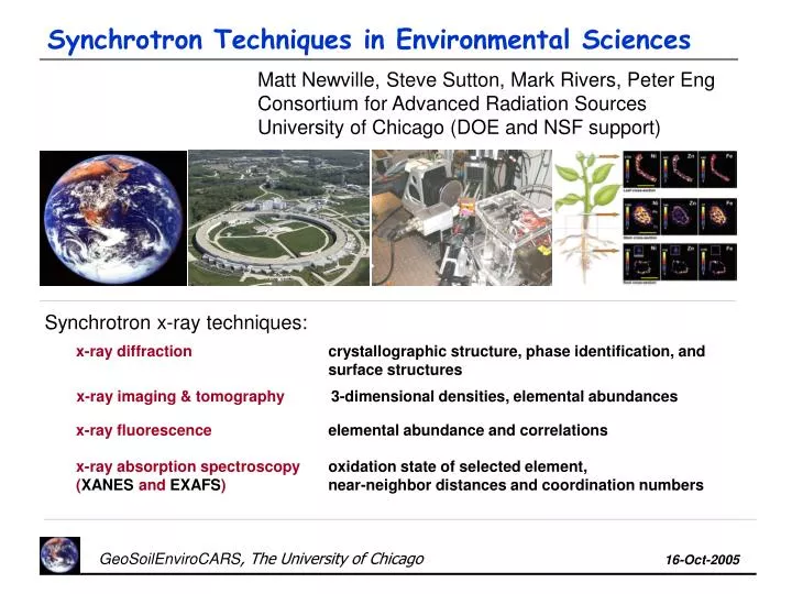
Matt Newville, Steve Sutton, Mark Rivers, Peter Eng Consortium for Advanced Radiation Sources University of Chicago (DOE and NSF support). Synchrotron Techniques in Environmental Sciences. Synchrotron x-ray techniques:. x-ray diffraction.

E N D
Matt Newville, Steve Sutton, Mark Rivers, Peter Eng Consortium for Advanced Radiation Sources University of Chicago (DOE and NSF support) Synchrotron Techniques in Environmental Sciences Synchrotron x-ray techniques: x-ray diffraction crystallographic structure, phase identification, and surface structures x-ray imaging & tomography 3-dimensional densities, elemental abundances x-ray fluorescence elemental abundance and correlations x-ray absorption spectroscopy (XANES and EXAFS) oxidation state of selected element, near-neighbor distances and coordination numbers
G. E. Brown, Jr. and N. Sturchio identified these important issues in low-temperature geochemistry and environmental science: From: An Overview of Synchrotron Applications in Low Temperature Geochemistry and Environmental Science. Reviews of Mineralogy & Geochemistry (vol 49, 2002): • In-situ studies, e.g. in the presence of water, water vapor, biota, … are critical. • Molecular-level speciation of trace environmental contaminants are necessary for understanding their behavior. • Complex natural systems and model systems must be studied in parallel. • Complementary characterization and modeling methods are necessary. • The nature of solid/water interface and sorbed species must be known. • Molecular mechanisms of bio- and phyto-remediation must be understood. X-ray Applications for Geo/Environmental Sciences Synchrotron x-ray techniques are powerful tools for addressing these issues.
What is a Synchrotron? A very bright x-ray source Advanced Photon Source, Argonne National Lab, Argonne Illinois 7 GeV electron storage ring producing high-brilliance x-ray beams. ~40 experimental stations running simultaneously, with a wide range of applications. 1 of 4 US DOE run x-ray sources operated as User Facilities (easy access). Many similar machines throughout the world. Electrons accelerated to 7GeV emit hard x-rays (1 to 100 keV).
Synchrotron: a very bright x-ray source Advanced Photon Source, Argonne National Lab, Argonne Illinois 7 GeV electron storage ring producing high-brilliance x-ray beams. ~40 experimental stations running simultaneously, with a wide range of applications. 1 of 4 US DOE run x-ray sources operated as User Facilities (easy access). Many similar machines throughout the world. GSECARS: 1 of ~7 stations doing Environmental Science at the APS
X-ray Properties of Synchrotrons x-ray brilliance for conventional laboratory and synchrotrons: brilliance = # of monochromatic x-rays per second, per area, per solid angle: how many monochromatic x-rays in a beam of light? Synchrotron x-rays have a broad energy spectrum “white light”, and are collimated in space. They can be focused to a few microns (sometimes smaller) or as large as several millimeters in size. x-rays are mostly non-destructive.
Typical Experimental Station: (x-ray microprobe) Sample Stage:x-y-z stage, 1mm resolution Incident Beam: LN2 cooled Si (111) mono CCD Camera:Bruker area detector Fluorescence detectors: 16-element Ge detector with DXP electronics Si-drift detector (shown) Lytle Ion Chamber Bent Laue Analyzer Wavelength Dispersive Spectrometer Optical Microscope: 5x to 50x objective with external video system Focusing:Kirkpatrick-Baez mirrors: Rh-coated Si, typically using 2x3mm spot sizes, at 50mm from end of mirrors. Entrance Slits:typically 250mm X 250mm, accepting ~30% of undulator beam
X-ray Diffraction / Scattering: Determine the crystallographic phases in a sample, study surface and interface structures Quantitative: very precise / accurate determination of crystalline phases X-ray Diffraction and Scattering Small Spot Size: x-ray beam sizes of a few microns make very small phases visible. Several modes available using synchrotron radiation: Single Crystal Diffraction (precise atomic positions) Powder diffraction (phase identification, unit cell refinement) Small / Wide Angle Scattering (nanometer-scale structure) Surface / Interface studies (surface structure)
X-ray Tomography: x-ray absorption radiography collected at different angles to look in the interior of objects High Resolution: micron-scale 3D volumes can be made of millimeter sized objects. X-ray Absorption Tomography See “inside” sample: without actually slicing precious or sensitive objects, one can make any “virtual slice” desired. Visible light Sample x-rays x-rays CCD camera Get beautiful images: can quickly aide understanding of system. w Phosphor Microscope objective rotation stage Get full 3d volume: can be put into mathematical models of fluid flow, pore volume connectivity, etc. Can get elemental specificity: by going above/below an absorption edge – works for elements at wt% level. Eocene age fossil
X-ray Fluorescence: characteristic x-ray emission lines from de-excitation of electronic core levels for each atom. Element Specific: All elements with Z>~14 are visible. It is usually easy to distinguish different elements. X-ray Fluorescence and Microprobe Quantitative: precise and accurate elemental abundances can be made. Low Concentration: concentrations down to ppm level can be seen. Natural Samples: samples can be in solution, liquids, amorphous solids, soils, plant roots, surfaces, etc. Small Spot Size: measurements can be made on samples down to a few microns in size. Combined with Other Techniques: XRD, XANES, EXAFS
X-ray Absorption Spectroscopy: energy-dependence x-ray absorption coefficient m(E) for a core-level electron of an element Element Specific: Elements with Z>14 can have EXAFS measured X-ray Absorption Spectroscopy: XANES and EXAFS Valence Probe: XANES is sensitive to chemical state and formal valence of selected element. Local Structure Probe: EXAFS gives atomic species, inter-atomic distance, and number of near-neighbor atoms around a selected element.. Low Concentration: ~10 ppm for XANES, ~100 ppm for EXAFS. Natural Samples: samples can be in solution, liquids, amorphous solids, soils, plant roots, surfaces, etc. Small Spot Size: XANES and EXAFS measurements can be made on samples down to ~5 microns in size. XANES = X-ray Absorption Near-Edge Spectroscopy EXAFS = Extended X-ray Absorption Fine-Structure
XANES: Oxidation State and Coordination Chemistry X-ray Absorption Near-Edge Spectroscopy (XANES) gives a direct measurement of chemical state and valence state of an element.. For atoms with partially filled d orbitals, the amount of p-d hybridization dramatically changes when the local coordination goes from octahedral to tetrahedral. This gives dramatic changes in XANES, including pre-edge peaks, which are due to unfilled d orbitals that can be filled by an s->p transition only with orbital hybridization. This depends strongly on coordination chemistry and formal oxidation state.
Nicola Allison, Adrian Finch (Univ of Brighton, Univ of Hertfordshire, UK) Strontium Paleothermometer in Coral: XRF The abundance of Sr in aragonite (CaCO3) formed by corals is used to estimate of seawater temperature and composition at formation time. [Sr]/[Ca] ~ T XRF MAPS of a section of the coral were made at 5mm resolution. Sr and Ca fluorescence (and other trace elements) were measured simultaneously at each pixel with a multi-element solid-state detector. The Sr and Ca maps show incomplete correlation and substantial variations in [Sr]/[Ca] on length scales consistent with a diurnal growth cycle. Ca Sr 200mm 300mm Sr XAFS was measured at a spot with high [Sr] -- above the solubility limit of Sr in aragonite. SEM images of Night growth (Left) and Daytime growth (Right)
Nicola Allison, Adrian Finch (Univ of Brighton, Univ of Hertfordshire, UK) Strontium Paleothermometer in Coral: EXAFS Since the Sr concentration was above its solubility limit (~1%) in aragonite, it was not known if Sr would precipitate out into strontianite (SrCO3: a structural analog of aragonite), or remain in the aragonite phase. First shell EXAFS is same for both strontianite and aragonite: 9 Sr-O bonds at ~2.5A, 6 Sr-C at ~3.0A. Second shell EXAFS clearly shows Sr-Ca (not Sr-Sr) dominating, as shown at left by contrast to SrCO3 data, and by comparison to simulated EXAFS spectrum of Sr substituted into aragonite. The coral traps Sr in thermodynamically-unfavorable aragonite structure, even at super-saturated concentrations.
Metal Uptake in Ni Hyperaccumulating Plants D. Sparks, D. McNear, E. Peltier , U. of Delaware How is Ni taken up, transported, and stored in the hyperaccumulating species Alyssum murale (mustard family)? Can we improve our basic understanding of phytoremediation, and hopefully optimize it? Samples were grown both hydroponically and in Ni enriched soils. Fluorescence Tomography: focused x-ray beam Sample Transmission detector fluoresced x-rays w Alyssum murale x Fluorescence tomography allows us to measure metal distribution in the interior of plant material without physically slicing the plant. rotation and translation stages fluorescence detector
X-ray Tomography Results for Alyssum Murale D. Sparks, D. McNear, E. Peltier, et al., U. of Delaware Leaf: Ni is in epidermal cells and veins but not in mesophyll cell. Zn is in the veins and exterior walls. Stem: Ni is in epidermis, pith and other ground tissues. The phloem side of vascular bundles has little Ni, the xylem is enriched in Ni. Zn is at the interface of the epidermis and vascular system. Root: Ni, Zn, and Fe are all present on root exterior in dried roots, and seen in the interior of wet roots. Virtual slices through alyssum murale grown in Ni-enriched soil
L K H Q sample P. Eng, S. Ghose (U. Chicago), T. Trainor (U Alaska, Fairbanks) Surface Scattering, the hematite surface Surface x-ray diffraction:A surface disrupts the infinite 3D lattice that make Bragg diffraction spots, and moves diffraction intensity to lines “between the Bragg points”. The q-dependence and shape of these crystal truncation rods is sensitive to the roughness and atomic arrangement at the crystal surface. Sample: single crystal wafer of (1-102) a-Fe2O3, 0.5mm thick, fully hydrated, clean, and then with 100mM Pb sorbed on the surface.
P. Eng, S. Ghose (U. Chicago), T. Trainor (U Alaska, Fairbanks) Surface Structure of Fe2O3: CTR Results CTR data for hydrated (1 -1 0 2) Fe2O3 surface with two structural models: Bulk termination Missing Fe termination The bulk termination gives a poor fit, and removing one Fe from the termination gives a much better match to the measurement.
100mM Pb(II) Unreacted Acid washed L (r.l.u.) P. Eng, S. Ghose (U. Chicago), T. Trainor (U Alaska, Fairbanks) Surface Structure of (1-102)Fe2O3 with 100mM Pb Pb(II) sorption isotherms on the hematite (1-102) surface: Facet-specific adsorption curves can be measured. CTR data gives the structure of the “ordered” adsorption complexes and fractional site occupancy Such experimental adsorption isotherms give facet-specific binding energies that can be compared to calculations.
G. E. Brown, Jr. and N. Sturchio identified these important issues in low-temperature geochemistry and environmental science: From: An Overview of Synchrotron Applications in Low Temperature Geochemistry and Environmental Science. Reviews of Mineralogy & Geochemistry (vol 49, 2002): • In-situ studies, e.g. in the presence of water, water vapor, biota, … are critical. • Molecular-level speciation of trace environmental contaminants are necessary for understanding their behavior. • Complex natural systems and model systems must be studied in parallel. • Complementary characterization and modeling methods are necessary. • The nature of solid/water interface and sorbed species must be known. • Molecular mechanisms of bio- and phyto-remediation must be understood. X-ray Applications for Geo/Environmental Sciences Synchrotron x-ray techniques are powerful tools for addressing these issues.