Download
1 / 55
550 likes | 554 Views
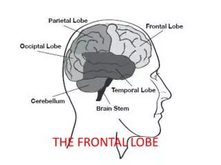
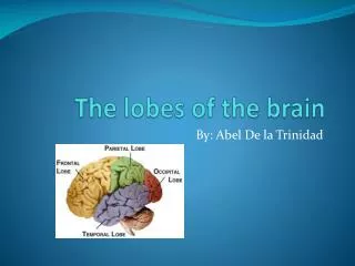
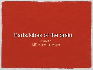
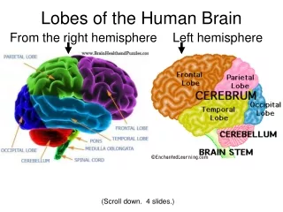
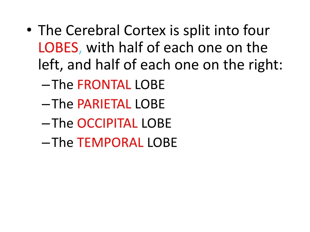
The Cerebral Cortex is split into four LOBES , with half of each one on the left, and half of each one on the right: The FRONTAL LOBE The PARIETAL LOBE The OCCIPITAL LOBE The TEMPORAL LOBE. The Frontal Lobes are the portions of the cortex lying just behind the forehead

E N D
The Cerebral Cortex is split into four LOBES, with half of each one on the left, and half of each one on the right: • The FRONTAL LOBE • The PARIETAL LOBE • The OCCIPITAL LOBE • The TEMPORAL LOBE
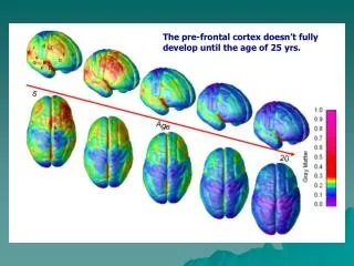
The Frontal Lobes are the portions of the cortex lying just behind the forehead • Mostly involved in abstract thought, speaking, muscle movements, making plans, and judgments
In the left frontal lobe specifically, there is an association area called Broca’s Area
Broca’s Area controls language expression and the muscle’s involved with producing speech
Along the top of both frontal lobes runs the motorcortex Sensory Motor Integration
The motor cortex sends messages back to the muscles of the body in order to control voluntary movements.
The Parietal Lobes are the portion of the cortex lying at the top of the head, and includes the sensory(or somato-sensory)cortex
The sensory cortex registers and processes touch sensations, temperature, pressure.
The parietal lobes play important roles in integrating sensory touch information, and in the handling and manipulation of objects.
The Temporal Lobes are the portions of the cerebral cortex roughly located above the ears • The functions of the temporal lobes are generally specific to audio processing, and may extend to comprehension, naming, verbal memory and other language functions.
In the left temporal lobe specifically, there is an association area called Wernicke’s Area
Wernicke’s Area interprets both written and spoken language.
Aphasia is a disorder of either Broca’s or Wernicke’s areas. • You may be able to comprehend, but cannot intelligibly communicate, or vice-versa.
Broca’s Aphasia - Broca's aphasia characterizes patients as people who have loss the production of complete sentence structures in speech and writing. • Wernicke’s Aphasia - Individuals with Wernicke's aphasia speak in long, uninterrupted sentences; however, the words used are frequently unnecessary or even made-up. They have a great deal of difficulty understanding other people's speech, sometimes to the point of being unable to understand spoken or written language at all.
The Occipital Lobes are located at the back of the head. • The occipital lobe is responsible for processing visual information.
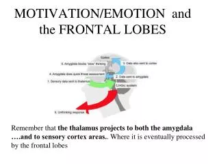
Collectively, the thalamus, hypothalamus, hippocampus, and the amygdala are known as the Limbic System as well
The THALAMUS receives sensory input from all of the senses except smell, and routes it to the proper area of the brain for processing
The HYPOTHALAMUS is responsible for several maintenance activities, including eating, drinking, body temperature, and sexual arousal
The HYPOTHALAMUS also relays communication between the brain and the endocrine system, via the pituitary gland, and then monitors the hormones released into the bloodstream
The HIPPOCAMPUS is essential to transferring short term memory to long term memory
The AMYGDALA: • Processes and recognizes emotions, especially those tied to anger, disgust, fear • Emotional aspects of memory stored here
II. The Midbrain • The Midbrain is located in the center of the brain and connects the forebrain to the hindbrain. It assists in: • Motor control • Hearing • Alertness and sleep/wake cycles • Temperature regulation
The RETICULAR FORMATION runs from the spine up and through the midbrain and connects to the thalamus. It is responsible for: • Visual tracking • Relaying audio and visual information to the cerebellum • Pain sensations • Attentiveness and consciousness • Selective attention
The hindbrainis the oldest and innermost region of the brain. It independently controls most life-sustaining functions of the body.
There are three brain areas associated with the Hindbrain: • Medulla • Cerebellum • Pons
The point at which the spinal cord enters the skull is called the MEDULLA. It is responsible for: • Involuntary functions such as breathing, heart rate and blood pressure.