Download
1 / 24
260 likes | 288 Views
CHAPTER 4 ISOLATION, QUANTIFICATION AND identification OF VIRUSES. REVISION. VIRULENT VIRUS - Upon entering the host, the virus circular DNA will undergo multiplication and lyses the host to release the new virion. The Lytic Cycle Culminates in the death of the host cell

E N D
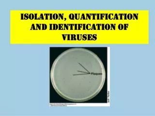
CHAPTER 4 ISOLATION, QUANTIFICATION AND identification OF VIRUSES
REVISION VIRULENT VIRUS - Upon entering the host, the virus circular DNA will undergo multiplication and lyses the host to release the new virion. • The Lytic Cycle • Culminates in the death of the host cell • Virulent viruses reproduce only by lytic cycle. • T4 virulent phages • The Lysogenic Cycle • Replication of the viral genome without destroying the host cell. • A temperate virus may reproduce by either cycle. • Lambda virus (temperate phage): resembles T4 but only has a single short tail fiber • TEMPERATE VIRUS • Within the host, the virus’ circular DNA engages in either the lytic or lysogenic cycle. • - During a lytic cycle, the viral genes immediately turn the host cell into a virus-producing factory, and the cell soon lyses and releases its viral products.
Regardless of the type of virus, the parasite diverts the host cell’s resources for viral production. • The host cell provides: • Nucleotides for nucleic acid production • Enzymes • Ribosomes • tRNA • Amino acids • ATP Machinery for protein synthesis
Phage Growth Growth curve for a bacteriophage: The eclipse phage represents the time after penetration through the biosynthesis of mature phages. The latent period represents the time after penetration through release of mature phages. The number of viruses per infected cell is the viral yield, or burst size
Lesson Outcome • Explain the cultivation and quantification techniques for bacteriophages
Cultivation and identification of viruses • The primary purposes of viral cultivation are: • 1. to isolate and identify viruses in clinical specimens • 2. to prepare viruses for vaccines • 3. to do detailed research on viral structure, multiplication cycles, genetics, and effects on host cells. • Bacteriophages – cultivation and identification is simple and easy, due to the simplicity of the host cells. • Animal viruses – difficult, due to the properties of the animal host. Systems of cultivation with broader applications were developed, including in vitro* cell (or tissue) culture methods and in vivo* inoculation of laboratory-bred animals and embryonic bird tissues. • - Such use of substitute host systems permits greater control, uniformity, and wide-scale harvesting of viruses.
Cultivation and identification of phages Obtaining bacteriophage from sample Amplification/multiplication of phages Isolation of multiplied phages Plaque assay Solution (sample) into liquid media (eg. NB, TSB) increase the numbers of phages (in the sewage sample) by allowing them to infect and reproduce within fresh host. Addition of host – sewage: enteric bacteria, faeces: E.coli Incubation: 37o C, 24 hrs Separate the remaining host cell/cell debris via centrifugation and filtration (0.2µm filter) Preparation of pure phage suspension Detection, identification , phage isolation for storage and future research
Isolation and identification of phages – Plaque assay technique • Plaque assay technique • STEPS: • Serial dilutions –ten-fold dilution in preparation of phage suspension • Add in host (log-phase growth) to phage dilution • Incubation 37o C, 20 min • Add in top agar • Pour on solidified agar • Incubate 37o C, 18-24 hrs. • Observation of plaque formation Detection, isolation, identification, characterisation of phages To allow infection of phage to host
Isolation and identification of phages – Plaque assay technique • Plaque assay technique • STEPS: • Serial dilutions –ten-fold dilution in preparation of phage suspension • Add in host (log-phase growth) to phage dilution • Incubation 37o C, 20 min • Add in top agar • Pour on solidified agar • Incubate 37o C, 18-24 hrs. • Observation of plaque • formation Plaque: can be collected for storage
Identification of phages – Plaque assay technique • Plaque ? • The basis is that one viral particle infects one cell, is replicated and the cell lyses. The nearby cells are infected and a ‘plaque’ of dead cells is formed over time. Zone of cell death/ a clear area in a bacterial lawn culture where viruses have lysed host cells HOW TO IDENTIFY TEMPERATE PHAGE? - Cloudy plaque
Identification of phages – Plaque assay technique • Basis of plaque formation: • Plaque assay – also to calculate number of phages present. • The titer of a phage suspension, is determined • by counting the number of plaques that form from a given • volume of suspension. Phage titer is expressed as plaque forming units (PFU) per milliliter (ml). • pfu/ml*measurement of the number of viable, infectious bacteriophage
QUIZ 1. List the replication steps for animal viruses. 2. Name the point of entry and exit for animal viruses. 3. Name the site for replication, protein synthesis and maturation step for DNA virus. 4. Define “plaque”. 5. How do you identify the present of lambda phage through plaque assay technique? Adsorption, Penetration, Uncoating, Synthesis, Maturation, Release Entry: endocytosis and fusion of virus envelope to host cell membrane Exit: budding/exocytosis and lysis Replication: nucleus Protein synthesis: cytoplasm Maturation: nucleus A clear area in a bacterial lawn culture where viruses have lyzed host cells Formation of cloudy/not clear plaque because lambda phage is temperate phage
Overview of Animal Viruses - Overview of animal virus actions
Lesson Outcome • Explain the cultivation and quantification techniques for animal viruses
Isolation, Cultivation and Identification of animal viruses CULTIVATION/ ISOLATION 1. In living animals - using liveanimal eg.mice, rats, rabbits, guinea pigs, hamster, chickens, and monkey. - the animal is exposed to the virus by injection of a viral preparation or specimen into the brain, blood, muscle, body cavity, skin, or footpads. - use in example research to study the immune system’s response to viral infections. - HIV: immunodeficient mice grafted to produce human T cells and human gamma globulin. - The signs of viral growth include death of the animal and defects in animal development. The infected animal tissue can be prepared for examination with an electron microscope IDENTIFICATION
Isolation, Cultivation and Identification of animal viruses 2. In Embryoted egg - use embryonated chicken, duck or turkey for inoculation of viral suspension. - The signs of viral growth include death of the embryo, defects in embryonic development, and localized areas of damage in the membranes, resulting in discrete, opaque spots called pocks (a variant of pox). The embryonic fluid and tissue can be prepared for examination with an electron microscope. - Some can also be detected by their ability to agglutinate red blood cells or by their reaction with an antibody of known specificity that will affix to its corresponding virus, if it is present. CULTIVATION/ ISOLATION IDENTIFICATION
Viral culture in eggs: Some viruses, such as influenza viruses, are grown in embryonated chicken eggs
3. Using cell culture - preferred type of growth medium for virus, more convenient than the previous two methods - use isolated cell from animal that are cultured invitro. Normal cells will form monolayer. - If viruses are present, the cells of monolayer will deteriorate as they multiply. Cell deterioration is called cytopathic effect (CPE). CPE can be detected and counted = plaques by phages (plaque assay). Microscopic observation via electron microscope (histopathology). CULTIVATION/ ISOLATION IDENTIFICATION A. Normal B. Transformed
Culturing of using cell culture • Two discoveries greatly enhanced the usefulness of cell cultures for virologists and scientists • The discovery and use of antibiotics made it possible to prevent bacterial contamination • The discovery of proteolytic enzymes (e.g. trypsin) can free animal cells from surrounding tissues without injuring freed cells • Subculturing: the process by which cells from an existing culture are transferred to new containers with fresh nutrient media
Identification of viruses 1. PCR – polymerase chain reaction 2. Restriction fragments polymorphisms (RFLP) 3. Serological method – Western blot common method use 4. Immunological test , ELISA, agglutination test – if specific antibody is available A preparation of killed, inactivated or attenuated microorganisms to induce artificially acquired active immunity Vaccine development Embryoted chicken egg – one the most used method of viral isolation and growth Still used to grow viruses for some vaccines – eg. Influenza vaccine Cell culture and animal tissue are also used in vaccine preparation for some viruses.
QUESTION • Briefly explain the culturing method used to identify, isolate and cultivate animal viruses. Embryonated eggs : use embryonated chicken, duck or turkey for inoculation of viral suspension. The signs of viral growth include death of the embryo, defects in embryonic development, and localized areas of damage in the membranes, resulting in discrete, opaque spots called pocks (a variant of pox). The embryonic fluid and tissue can be prepared for examination with an electron microscope. Tissue culture: use isolated cell from animal or plant that are cultured invitro. The cells will form monolayer. Thesign of viral growth detected through formation of plaque or looking at cytopathic effect. Animal : using live animal eg. mice, rats, rabbits, guinea pigs, hamster, chickens, and monkey. The signs of viral growth include death of the animal and defects in animal development. The infected animal tissue can be prepared for examination with an electron microscope. - Identification: also by PCR, serology