Download
1 / 20
200 likes | 227 Views
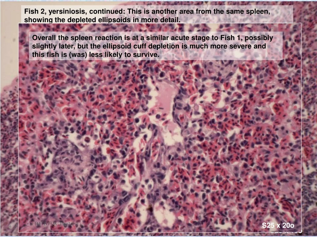
Fish 2, yersiniosis, continued: This is another area from the same spleen, showing the depleted ellipsoids in more detail.

E N D
Fish 2, yersiniosis, continued: This is another area from the same spleen, showing the depleted ellipsoids in more detail. Overall the spleen reaction is at a similar acute stage to Fish 1, possibly slightly later, but the ellipsoid cuff depletion is much more severe and this fish is (was) less likely to survive. S25 x 20o S25 x 10o
Fish 3, yersiniosis: This spleen shows a more obvious lymphoid response. (But don’t overinterpret this as an indicator of the stage of response to any specific pathogen - it could relate to antigens encountered before invasion of the current pathogen). The ellpsoids show enlarged pale cuffs. Higher magnification shows much more adhesion of the macrophages making up the primary cuff, with less open oedematous spaces and migrating cells - signs of a later ellipsoid response. Some outer peripheral dark cells are present, and the spleen appears robust though active. The well developed ellipsoid response adds weight to the likelihood that the lymphoid reaction is a response to this pathogen. Overall the appearance is of a late stage of what appears to be an effective reaction to this pathogen, and suggests this fish had a reasonable chance of surviving this infection. S26 x 4o S26 x 20o
Pigmentation and the spleen We have seen several examples of individual pigmented macrophages, containing the residual pigments of phagocytosis and destruction of pathogens or damaged cells. We noted that these generally migrate to form melanomacrophage centres, though this reaction is slight in salmonids. More generalised pigmentation in the spleen if generally derived from erythrocytes, through: • as artefact (particularly during slow fixation) • erythrocyte lysis (often post-haemorrhage) • erythrophagocytosis (of intact but damaged red cells)
Spleen pigment 1: melanomacrophage centres Recap: • A variety of pigment products are formed in macrophages after phagocytosis and destruction of pathogens or damaged host cells. These cells later migrate to form melanomacrophage centres. • The extent of migration / aggregation, and the colour of pigment varies between the species. (We have seen some examples). • As the nature of the phagocytosed material also varies with the nature of the incident, the proportion of specific pigments (such as iron containing pigments, which are recycled), will also vary.
This Goldfish spleen shows an area near a major vessel with aggregations of the slightly pigmented pale melanomacrophages (MMs) typical of this species (eg centre right). Perl’s stain for iron demonstrates a little iron positive pigment (dark blue) within this MM centre. The stored iron is derived from haemoglobin. The amount of pigment generally reflects the level of erythrocyte turnover and haematopoiesis in this organ (and will also reflect species variation in the sites of haematopoiesis). S26 x 20o
Just for fun, here is the aged (and autolysed) Orego Dory in poor condition and with a heavy parasite load, that we looked at in a kidney presentation. Note the extent of melanomacrophage centre formation, as well as the large autolysed ellipsoid cuffs. (And there is a metazoan parasite to boot.) This fish had an ongoing poor energy balance, with high levels of tissue turnover, probably some of this parasite driven. x 4o
What do you think is the nature of the pigment in this slide? Note there are actually 2 types of pigment to consider. The heavily pigmented cells within the ellipsoid cuffs appear likely to be melanomacrophages. But note that there is also pigment within the free flowing blood. One of the common findings after formalin fixation of in any organ containing abundent blood (especially where fixation is slow or the fixative suboptimal), is the formation of so-called “formalin pigment” within the blood. This must be recognised as an artifact and not confused with the deposition of pigment within macrophages (which may show 2 patterns). x 40o x 20o x 10o
Macrophage pigment, pattern 1: The spleen from this Atlantic salmon (which was recovering from exposure to the toxin of Aurelia sp. Jellyfish) shows fine pigmentation round an ellipsoid. Note the location - the pigment forms a ring approximately 1 cell from the fenestrated endothelium. S23 x 40o
This is a low power view of the same spleen. Note the distribution pattern of the pigment - it occupies a similar position in a large number of the ellipsoid cuffs. What do you think is the origin of this pigment? Pigment along an ellipsoid capillary in longitudinal section. The fine nature of the pigment, and the relatively uniform distribution within a band of macrophages adjacent to the ellipsoid are indication that this is small fragments of red cells and / or free haem pigment from ruptured erythrocytes, such as will occur following a heamorrhage into the tissues. The relatively uniform band close to the ellipsoid vessel suggest a sudden recent episode of haemorrhage. This has probably occurred within the last day or 2, although the temperature will affect metabolic rate and therefore generally prevent a more exact time estimate. S23 x 40o S23 x 20o S23 x 10o
One of the most obvious changes is the amount of pigment. This is present within melanomacrophages which are aggregated into MM centres to an extent that is unusual in salmonids. Note these cells have migrated away from the ellipsoid cuffs (circled). Note the unusual “muddy” color of many erythrocytes migrating through the cuffs, and in the interstitium, plus the pale cells. This small (50g) rainbow trout shows a dense spleen with an architecture reflecting the ellipsoid pattern but with large pale areas showing little resemblance to the normal ellipsoid pattern, much dense interstitial tissue. The latter alone is an indication of a quite prolonged time course. [In fact this sample was taken 21 days after a problem in this tank was first noted.] One of the most obvious changes is the amount of pigment. This is present within melanomacrophages which are aggregated into MM centres to an extent that is unusual in salmonids. These contain pigment of varying density – which is consistent with the clinical history of an ongoing problem of several weeks duration (the pale pigment being the most recent). S30 x 4o S28 x 20o S30 x 10o
Red cell changes are more clearly seen in this fish which was was sampled earlier in the same episode (1 week after problem first noted). Anemia and blood cell abnormalities (which we will return to in the next presentation) were present. Pigmented melanomacrophages are abundant, but most contain pale pigment (as expected from time-frame). Note the changed color of the marked erythrocytes ( white arrows) ), and the apparent vacuoles in others (blue arrows). S001559 x 40o
Erythrophagocytosis & erythrocyte damage. • The preceding slides were from an episode of nitrite toxicity. Note the duration – nitrite toxicity in mammals tends to be an acutely fatal toxicity of dietary origin, typically occurring suddenly after movement of stock to pastures with high nitrite levels. Clinically there is formation of sufficient methaemoglobin to give visibly brown blood, stopping oxygen transport. Methaemoglobin is formed by the strong oxidising action of nitrite. • Such acute reactions, and visibly brown blood, can occur in fish, but exposure is generally from elevated water nitrate rather than dietary origin, and is more likely to occur gradually. • This exposure pattern usually results in lower levels of exposure, with the cells of the gill, and the blood flowing through the gill being the most exposed tissues. Only small areas of haemoglobin may be damaged, which may not be sufficient for a visible change to blood colour, but which may be recognised by the spleen as damaged erythrocytes and removed. Other components such as cell membranes may be similarly oxidised. Gill epithelia may be damaged. • The pathology and pathogenesis is essentially similar for other strong oxidising agents present in water. • Note also secondary toxicity may occur – other toxicants can damage biofilter organisms, so nitrite produced is no longer removed.So always test nitrite too.
The next 2 fish are more examples from the same cohort. Note the similar pattern of melanomacrophage centres and pale vacuolated macrophages. Note the similarities in distribution of the pigment, the organising interstitial areas (indicating a sub-acute timeframe), and the “muddy” appearance of some of the abnormal blood cells, reflecting the changes in haemoglobin. Note that the altered haemoglobin staining is generally strongest in phagocytosed cells: initial damage may include alteration of haemoglobin, but this process also occurs after phagocytosis, as the haemoglobin is broken down by the phagocytes. S28 x 4o S28 x 10o S28 x 40o S28 x 20o
This fish (same cohort) shows more interstitial reaction, confirming a prolonged insult. Note the abundent pigment …….. At high magnification can readily see the most altered haemoglobin present in phagocytosed erythrocytes. S29 x 40o S29 x 4o S29 x 10o S29 x 20o
Another salmonid (rainbow trout)with similar pathology – but this was exposed to high levels of chlorine (another strong oxidising agent) rather than nitrite. Note pigment and large numbers of eythrocytes with altered haemoglobin, some of which are clearly in vessels or phagocytes. S X x 40o S (041259-5) x 10o S X x 20o
4 views of damaged erythrocytes with similar “muddy” haemoglobin, some phagocytosed, in the kidney interstitial tissue of another Atlantic salmon case. (The damaged erythrocytes were less obvious in the spleen, but may be phagocytosis in either organ.) In this case high H2S levels (rising from below the cage) is the suspected oxidising agent.
Focal spleen reactions& disturbances of architecture Focal reactions of the spleen may be caused by: • focal insults • viral diseases • contained or partially contained bacterial diseases (post septicaemia) • granulomatous (contained) bacterial and parasitic disease
Focal spleen reactions & disturbances of architecture 1. Focal insults.