Download
1 / 46
540 likes | 625 Views
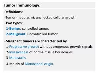
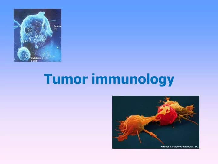
Tumor immunology. Tumor antigens. Tumor – specific antigens (TSA) complexes of MHC gp I with abnormal fragments of cellular proteins ( chemically induced tumors , leukemia with chromosomal translocation )

E N D
Tumor antigens • Tumor – specificantigens (TSA) • complexesof MHCgp I withabnormalfragmentsofcellularproteins(chemicallyinducedtumors, leukemiawithchromosomaltranslocation) • complexes of MHCgpwithfragmentsofoncogenicvirusesproteins(tumorscaused by viruses: EBV, SV40, polyomavirus…) • abnormalformsofglycoproteins(sialylationofsurfaceproteinsof tumor cells) • idiotypesofmyeloma and lymphoma(clonotyping TCR and BCR)
Tumor antigens • b) Tumor - associated antigens (TAA) • present also on normal cells • differences in quantity, time and local expression • auxiliary diagnostic markers
Tumor - associated antigens (TAA) • onkofetalantigens -on normalembryoniccells and some tumor cells • -fetoprotein (AFP) - hepatom • carcinoembryonic antigen (CEA) - coloncancer • melanomaantigens - MAGE-1, Melan-A
Tumor - associated antigens (TAA) • antigen HER2/neureceptor for epithelial growth factor, mammary carcinoma • EPCAM – epithelial cell adhesion molecule, metastases • differentiation antigens of leukemic cells - present on normal cells of leukocytes linage • CALLA -acute lymphoblastic leukemia (CD10 pre-B cells)
Anti-tumor immune mechanisms If tumor cells are detected, in defense may be involved non-specific mechanisms(neutrophilic granulocytes, macrophages, NK cells, complement) and antigen-specific mechanisms (TH1 and TC cells, antibodies).
Anti – tumor defense • tumor cells are weakly immunogenic • regulatory T cells promote progression of cancer • occurs when tumor antigens are presented to T cells by dendritic cells activated in the inflammatory environment
Anti – tumor defense • DC are necessaryforactivationof antigen specificmechanisms • predominance ofTH1(IFN , TNF) • specific cell-mediatedcytotoxicreactivity –TC • activationof TH2 → stimulationof B cells→ tumor specificantibodiesproduction (involved in the ADCC) • tumor cells are destroyed by cytotoxicNK cells(ADCC) • interferons - antiproliferative, cytotoxiceffect on tumor cells - INF - DC maturation
Mechanisms of tumor resistance to the immune system • high variability of tumor cells • lowexpressionof tumor antigens • sialylation • someanticancersubstanceshave a stimulatingeffect • productionoffactorsinactivating T lymphocytes • expressionofFasL → T lymphocyteapoptosis • inhibitionofthefunctionordurabilitydendriticcells (NO, IL-10, TGF-)
Transplantation • = transfer oftissueor organ • autologous - donor = recipient • syngeneic- geneticallyidentical donor and recipient (identicaltwins) • allogeneic - geneticallynonidentical donor ofthesame species • xenogenic - the donor ofanother species
Allotransplantation • differences in donor-recipient MHC gp and secondaryhistocompatibilityAg • alloreactivityof T lymphocytes- the risk ofrejection and graft-versus-host disease
Tests prior to transplantation • ABO compatibility (matching blood group) -risk of hyperacute rejection (= formation of Ab against A or B Ag on graft vascular endothelium) • HLA typing (matching tissue type)-determining of HLA alelic forms by phenotyping or genotyping • Cross-match - detection of preformed alloantibodies(after blood transfusions, transplantation, repeated childbirth) • Mixed lymphocyte reaction - testing of T lymphocytesalloreactivity
HLA typing= determminationof HLA antigens on thesurfaceoflymphocytes • Carry out during the testing before transplantation and in determination of paternity • serotyping • genotyping
HLA typing 1) Serotyping (microlymfocytotoxic test)
HLA typing 2) Molecular genetic methods- genotyping 2a) PCR-SSP 2b) PCR-SSO 2c) PCR-SBT
Cross-match testing • determination of preformed alloantibodies • recipient serum + donor lymphocytes + rabbit complement → if cytotoxic Ab against donor HLA Ag are present in recipient serum , Ab activate complement → lysis of donor lymphocytes. Dye penetration into lysis cells. • positive test = the presence of preformed Ab → risk of hyperacute rejection! → contraindication to transplantation
Mixed lymphocyte reaction (MLR) Two – way MRL • determination of T lymphocytesalloreactivity • mixed donor and recipient lymphocytes → T lymphocytes after recognition of allogeneic MHC gp activate and proliferate • this assay was used to study possible donor - recipient incompatibilities for graft transplants to help predict better outcomes
One-way MRL • determination of recipient T lymphocytesreactivity against donor cells • donor cells treated with chemotherapy or irradiated lose the ability of proliferation
Rejection • hyperacute • accelerated • acute • chronic
Rejection • Hyperacute • Accelerated • Acute • Chronic
Hyperacute rejection • minutes to hours after transplantation • humoral mediated immune response mechanism: • if in recipients blood are present preformed or natural Ab(IgM anti- carbohydrate Ag) before transplantation→ Ab + Ag of graft (MHC gp or endothelial Ag) → graft damage by activated complement • the graft endothelium: activation of coagulation factors and platelets, formation thrombi, accumulation of neutrophil granulocytes prevention: • negative cross match before transplantation, ABO compatibility
Acceleratedrejection • 3 to 5 daysaftertransplantation • caused by reactivation of T lymphocytes (secondary response)
Acute rejection • days to weeks after the transplantation or after a lack of immunosuppressive treatment • cell-mediated immune response mechanism: • reaction of recipient TH1 and TC cells against Ag of graft tissue • infiltration by lymphocytes, monocytes, granulocytes around smallvessels → destruction of tissue transplant
Chronic rejection • from 2 months after transplantation • the most common cause of graft failure mechanism is not fully understood: • non-immunological factors (tissue ischemia) and TH2 response with production alloantibodies, pathogenetic role of cytokines and growth factors (TGFβ) • fibrosis of the internal blood vessels of the transplanted tissue, endothelial damage →impaired perfusion of graft → gradual loss of its function • dominating findings: vascular damage
Rejection • Factors: • Thegeneticdifferencebetween donor and recipient, especially in thegenescodingfor MHC gp (HLA) • Type oftissue / organ - thestrongestreactionsagainstvascularizedtissuescontaining many APC (skin) • Theactivityofthe recipientimmunesystem– theimmunodeficiency recipient has a smallerrejectionreaction; immunosuppressivetherapyaftertransplantation – suppressionofrejection • Status oftransplanted organ - thelengthofischemia, themethodofpreservation, traumatization of organ at collection
Graft-versus-host (GvH) disease • after bone marrow transplantation • GvH also after blood transfusion to immunodeficiency recipients • T-lymphocytes in the graft bone marrow recognize recipient tissue Ag as foreign (alloreactivity)
Acute GvH disease • days to weeks after the transplantation of stem cells • damage of liver, skin and intestinal mucosa • prevention: appropriate donor selection, the removal of T lymphocytes from the graft and effective immunosuppression
Chonic GvH disease • months to years after transplantation • infiltration of tissues and organs by TH2 lymphocytes, production of alloantibodies and cytokines → fibrosis • process like autoimmune disease: vasculitis, scleroderma, sicca-syndrome • chronic inflammation of blood vessels, skin, internal organs and glands, which leads to fibrosis, blood circulation disorders and loss of function
Graft versus leukemia effect (GvL) • donor T lymphocytes react against residual leukemick cells of recipient (setpoint response) • mechanism is consistent with acute GvH • associated with a certain degree of GvH (adverse reactions)
Immunopathological reactions • Immune response which caused damage to the body (Consequence of immune response against pathogens, inappropriate responses to harmless antigens; autoimmunity)
Immunopathological reactions Classification by Coombs and Gell IV types of immunopathological reactions: Type I reaction - response based on IgE antibodies Type II reaction - response based on antibodies, IgG and IgM Type III reaction- response based on the formation of immune complexes Type IV reaction - cell-mediated response
Immunopathological reaction type II (cytotoxic hypersensitivity reactions) • Cytotoxic antibodies IgG and IgM bind to cell surface antigens on own cells: • complement activation • binding to Fc receptors on phagocytes and NK cells (ADCC)
Examples of immunopathological reaction type II • Transfusionreactionsafteradministrationofincompatibileblood: bindingofantibodies to antigens on erythrocytes → activationoftheclassicalpathwayofcomplement → cell lysis • Hemolyticdiseaseofnewborns: caused by antibodiesagainstRhD antigen
Examples of immunopathological reaction type II Autoimmune diseases: • organ-specific cytotoxic antibodies (antibodies against erythrocytes, neutrophils, thrombocytes, glomerular basement membrane ...) • blocking or stimulating antibodies Graves - Basedow's disease - stimulating antibodies against the receptor for TSH Myasthenia gravis - blocking of acetylcholin receptor→ blocking of neuromuscular transmission Pernicious anemia - blocking the absorption of vitamin B12 Antiphospholipid syndrome - antibodies against fosfolipids Fertility disorder - antibodies against sperms or oocytes
Immunopathological reaction type III(immune-complex reactions) • circulating antigen- IgG antibody immune complexes that depositin tissues • immunocomplexes - activate complement - bind to Fc receptors on phagocytes • immune complexes, depending on the quantity and structure, are eliminated by phagocytes or stored in tissues
Immunopathological reactions type III • pathologicalimmunocomplexes response ariseswhenis a large dose of antigen, or antigen in the body remains; arise 10-14 daysafteraplicationofAg and inducedinflamation (canget to chronicstate) • immunecomplexes are deposited in thekidneys (glomerulonephritis), on thesurfaceofendothelialcells (vasculitis) and in synovie joint (arthritis)
Serum sickness • the therapeutic application of xenogeneic serum (antiserum to snake venom) • creation of immune complexes and their storage in the vessel walls of different organs • clinical manifestations: urticaria, arthralgia, myalgia Systemic lupus erythematosus • antibodies against nuclear antigens, ANA, anti-dsDNA Farmer's lung • IgG antibody against inhaled antigens (molds, hay) Post-streptococcal glomerulonephritis, cryoglobulinemia, revmatoid arthritis, post-infectious arthritis
Immunopathological reaction type IV(delayed-type reaction - DTH) • local reaction caused by TH1 cells and monocytes/macrophages (physiologically –defense against intracellular pathogens) • immunization by antigen → formation of antigen specific TH1 cells and memory cells • 12-48 hours after next antigen exposure arise local reaction → granuloma(TH1and macrophage infiltration) Tuberculin skin tes reaction Tissue damage in tuberculosis and leprosy Sarcoidosis Multiple sclerosis
Subtype IV - Cellular cytotoxic response(Tc activation) • similar to DTH reaction • TH1 cells activate CD8 + T lymphocytes • viral rashes • viral hepatitis • acute rejection of transplanted organ • some autoimmune thyroiditis • contact dermatitis
Contact dermatitis • is a localizedrashorirritationofthe skin caused by contactwith alergen (nickel, chromium, ingredients in cosmeticproducts, plant allergens and other) • thefirstissenzitization • appears in 24 – 48 hoursafter second contactwith alergen • diagnosis : patch test
Patch test • patch testis a methodused to determineif a specific substance causesallergicinflamationofthe skin • Allergens are applied to specialhypoallergenic patch on theback skin • Results are evaluatedafter48 and 72 hours • In positive reactionappearseczema
Tumourimmunology and immunotherapy https://www.youtube.com/watch?v=K09xzIQ8zsg • Thisishowyourimmunesystemfightscancer • https://www.youtube.com/watch?v=UM2f-qFZV3o