Download
1 / 25
440 likes | 848 Views
1962: Nobel Prize in Physiology and Medicine. James D. Watson. Francis H. Crick. Maurice H. F. Wilkins. What about? Rosalind Franklin. Nucleotides are composed of:. Sugar - deoxyribose. Phosphate Base - one of four types: adenine (A), thymine (T)

E N D
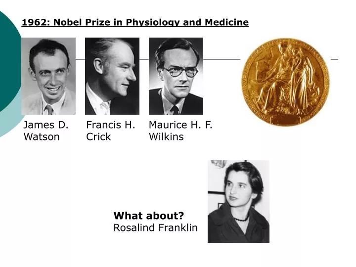
1962: Nobel Prize in Physiology and Medicine James D. Watson Francis H. Crick Maurice H. F. Wilkins What about? Rosalind Franklin
Nucleotides are composed of: Sugar - deoxyribose Phosphate Base - one of four types: adenine (A), thymine (T) guanine (G), cytosine (C) DNA (deoxyribonucleic acid) is a chain of nucleotides
Chemical Structure of DNA • Carbon 1 (C1) is where the base is attached. • Carbon 2 (C2) tells you if it is a ribose or deoxyribose. In deoxyribose "de oxy" is missing. • Carbon 3 (C3) is the point of attachment for more nucleotides. • Carbon 4 (C4) completes the ring via an oxygen which bridges to the carbon 1 (C1). • Carbon 5 (C5) hangs away from the ring and is the point of attachment for its phosphate(s).
The stands are "antiparallel," meaning they run in opposite directions. • Adenine (A) and thymine (T) form two hydrogen bonds between them. • Guanine (G) and cytosine (C) form three hydrogen bonds between them.
hydrogen bonds 5’ 3’ phosphodiester bonds 3’ 5’
A Typical Electrophoresis Gel Setup Negative end- - - - - - - - - - - - - Direction of DNA movement Direction of DNA movement Positive end+ + + + + + + + + +
Gel Electrophoresis • The gel acts as a sieve to filter the DNA fragments • Smaller fragments move faster through the gel than larger fragments
+ + + + + + + + + + + + + + + + + + + + + + + + ++ + + + + + + + + + DNA is negatively charged and therefore repelled from the negative pole and attracted towards the positive pole - - - - - - - - - - - - - - - - - - - - - - - - - - - - - - - - - - -
+ + + + + + + + + + + + + _ _ _ _ _ _ _ _ _ _ _ _
A Typical Image of an Agarose Gel Under UV Light Largest DNA fragments Decreasing DNA Size • The DNA fragments can be visualized using a special dye that specifically binds DNA and fluoresces under illumination with UV light Smallest DNA fragments
Sizing The Fragments of DNA • The sizes of the various fragments can be identified by including a “ladder” in the gel • A ladder is a mixture of DNA fragments of known size • When run in a gel electrophoresis, these fragments will separate into distinct bands that can be used as references
Typical Ladders-100 bp & 1 kbp (1000 bp) • The 100 bp ladder is composed of a mixture of small fragments (100 to 2000 bp) • The 1000 bp ladder is composed of a mixture of larger fragments (500 to 12000 bp)
Sizing a Gel Product Base Pairs (bp) 4000 3000 2000 1600 1000 500 2000 bp 1000 bp 1Kbp Sample ladder
What is the Size of this Fragment? 4000 3000 2000 1600 1000 500 Sample 1Kbp ladder
Background About Agarose • Agarose is a mixture of long chains of saccharides (sugar) • It is extracted from a seaweed and is used in oriental cuisine as a gelling agent for desserts • The greater the agarose concentration, the smaller the pores within the gel • This changes the ease with which the DNA can travel through the gel
Effects of Changing Gel Concentration • By increasing the agarose concentration the smaller DNA fragments will give a clearer separation • By lowering the agarose concentration the larger fragments of DNA will give a clearer separation • By optimizing the % agarose one can clearly separate a mixture of similar DNA fragments
Effect of Percent Agarose on Fragment SeparationAn example The 1kbp ladder and the 100bp ladder were run with 0.8 and 1.6 % agarose gels under the same conditions 1600 1000 1600 bp 500 400bp 1000 bp 300 bp 500 bp 200 bp 0.8% Agarose 80V – 2 hours 1.6% Agarose 80V – 2 hours Note how the bands have moved further in the low % agarose gel and the higher bp fragment are better defined. The low bp fragments are better defined in the high % agarose gel and are unresolved in the low % agarose gel
Purpose • Dye mixtures will be used to simulate DNA fingerprints. • Analyze the fragments produced from the different dyes.
Procedure for Day 1 • Determine amount of agar and buffer needed to make 50 ml of 1% agar. • Microwave the mixture for about 30 seconds. • Using a hot pad, take the flask out of the microwave and swirl gently. Make sure all the powder has dissolved. • Set on your bench to cool for 5 minutes. • Insert the comb into the first slot on the gel tray. • Pour the cool to the touch liquid gel into the tray. • Let the gel harden for at least 30 minutes. • Get a ziplock bag and write your group number on it. Leave the bag on the counter next to the gel.
Procedure for Day 2 • Pull out the comb from the wells by pulling gently upwards. • Place the gel into the gel box. Make sure the wells are at the black end. • Add 350 ml of TAE buffer to flood the gel and fill the wells. • Load 7 µl of each sample into a well on the gel. • During the lab, note where each sample was loaded on a diagram of your gel.
Put the cover on the electrophoresis box so that the electrodes are connected. Double check that the samples are closest to the negative (BLACK) electrode. • Plug the other end of the electrodes into the power supply and run at 100 volts for 30 minutes. • After the samples have run out, make a drawing of how the gel looked in your lab notebook.
Conclusion • What is the charge of these colored dyes? • What would happen to the samples if the gel were flipped around so that the sample wells were put closest to the positive (red) electrode, instead of the negative (black) electrode? • What would happen to the samples if you ran the gel at 200 volts for 15 minutes? • What is the charge on DNA?
Which colored dye moved the farthest distance through the gel? • Which colored dye moved the shortest distance through the gel? • Which colored dye is the smallest in size? • Which colored dye is the largest in size? • What is the relationship between distance traveled and size of the molecule in an agarose gel?