Download
1 / 49
490 likes | 495 Views
Investigation 9 BIOTECHNOLOGY: RESTRICTION ENZYME ANALYSIS OF DNA* How can we use genetic information to identify and profile individuals?. Pioneering DNA Forensics.

E N D
Investigation 9 BIOTECHNOLOGY:RESTRICTION ENZYME ANALYSIS OF DNA*How can we use genetic information to identify and profile individuals? Pioneering DNA Forensics file:///C:/Users/Andrew/Desktop/New%20AP%20Biology/AP%20Biology%20Labs/Inv.%208-%20Biotechnology-Bacterial%20transformation/Pioneering%20DNA%20Forensics%20%20%20NPR.htm
Learning targets for Restriction Digestion and Analysis of Lambda DNA lab? • Understand the use of restriction enzymes as biotechnology tools • Become familiar with principals and techniques of agarose gel electrophoresis • Generate a standard curve from a series of DNA size fragments • Estimate DNA fragment sizes from agarose gel data
learn how to use restriction enzymes and gel electrophoresis to create genetic profiles. • use these profiles to help Marcus and Laurel narrow the list of suspects in the disappearance of Ms. Mason.
Forensic DNA Fingerprinting: Using Restriction Enzymes
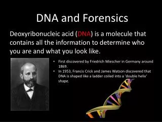
BACKGROUND-page 112 • Applications of DNA profiling extend beyond what we see on television crime shows. • Are you sure that the hamburger you recently ate at the local fast-food restaurant was actually made from pure beef? DNA typing has revealed that often “hamburger” meat is a mixture of pork and other non beef meats, and some fast-food chains admit to adding soybeans to their “meat” products as protein fillers. In addition to confirming what you ate for lunch. • DNA technology can be used to determine paternity, diagnose an inherited illness, and solve historical mysteries, such as the identity of the formerly anonymous individual buried at the Tomb of the Unknown Soldier in Washington, D.C.
DNA testing also makes it possible to profile ourselves genetically — which raises questions, including Who owns your DNA and the information it carries? This is not just a hypothetical question. The fate of dozens of companies, hundreds of patents, and billions of dollars’ worth of research and development money depend on the answer. • Biotechnology makes it possible for humans to engineer heritable changes in DNA, and this investigation provides an opportunity for you to explore the ethical, social, and medical issues surrounding the manipulation of genetic information.
DNA Fingerprinting Real WorldApplications • Crime scene • Human relatedness • Paternity • Animal relatedness • Anthropology studies • Disease-causing organisms • Food identification • Human remains • Monitoring transplants
The Forensic DNA Fingerprinting concepts: • DNA structure • DNA restriction analysis (RFLP) • Agarose gel electrophoresis • Molecular weight determination • Simulation of DNA Fingerprinting • Plasmid mapping
Lab Time Line • Restriction digest of DNA samples • Introduction to DNA Fingerprinting and RFLP analysis • Electrophoresis on Agarose gels • Analysis and interpretation of results
What is needed for restriction digestion? • Template DNA, uncut DNA, often bacterial phage DNA • DNA standard or marker, a restriction enzyme of known fragment sizes • Restriction enzyme(s), to cut template DNA • Restriction Buffer, to provide optimal conditions for digestion
The DNA DigestionReaction Restriction Buffer provides optimal conditions • NaCI provides the correct ionic strength • Tris-HCI provides the proper pH • Mg2+ is an enzyme co-factor
DNA DigestionTemperature Why incubate at 37°C? • Body temperature is optimal for these and most other enzymes What happens if the temperature is too hot or cool? • Too hot = enzyme may be denatured (killed) • Too cool = enzyme activity lowered, requiring longer digestion time
DNA Restriction Enzymes • Evolved by bacteria to protect against viral DNA infection • Endonucleases = cleave within DNA strands • Over 3,000 known enzymes
How does it work? • Enzyme Site Recognition • Each enzyme digests (cuts) DNA at a specific sequence restriction site • Enzymes recognize 4-, 6- or 8- base pair, palindromic sequences • Isoschizomers recognize identical sequences, but have different optimum reaction conditions and stabilities • Can be unambiguous or ambiguous Unambiguous Ambiguous
5 vs 3 Prime Overhang Enzyme cuts • Generates 5 prime overhang
5’GAATTC3’ 3’CTTAAG5’ 5’AAGCTT3’ 3’TTCGAA5’ 5’CTGCAG3’ 3’CACGTC5’ Common Restriction Enzymes • EcoRI • Escherichiacoli • 5’ overhang • HindIII • Haemophilus influensae • 5’ overhang • PstI • Providenciastuartii • 3’ overhang
Enzyme Site Recognition Restriction site Palindrome • Each enzyme digests (cuts) DNA at a specific sequence = restriction site • Enzymes recognize 4- or 6- base pair, palindromic sequences (eg GAATTC) Fragment 2 Fragment 1
5’GAATTC3’ 3’CTTAAG5’ ■■Activity I: Restriction Enzymes Read intro to activity 1. 1. What is the sequence of the complementary DNA strand? Draw it directly below the strand. • 5’-AAAGTCGCTGGAATTCACTGCATCGAATTCCCGGGGCTATATATGGAATTCGA-3’ • 3’-TTTCAGCGACCTTAAGTGACGTAGCGTAAGGGCCCCGATATATACCTTAAGCT-5’ 2. Assume you cut this fragment with the restriction enzyme EcoRI. The restriction site for EcoRI is 5’-GAATTC-3’, and the enzyme makes a staggered (“sticky end”) cut between G and A on both strands of the DNA molecule. Based on this information, draw an illustration showing how the DNA fragment is cut by EcoRI and the resulting products. • 5’-AAAGTCGCTGGAATTCACTGCATCGAATTCCCGGGGCTATATATGG AATTCGA-3’ • 3’-TTTCAGCGACCTTAAGTGACGTAGCGTAAGGGCCCCGATATATACCTTAAGCT-5’
PstI EcoRI Restriction Fragment Length PolymorphismRFLP GAATTC GTTAAC CTGCAG GAGCTC Allele 1 1 2 3 CGGCAG GCGCTC GAATTC GTTAAC Allele 2 3 Fragment 1+2 Different Base Pairs No restriction site M A-1 A-2 Electrophoresis of restriction fragments M: Marker A-1: Allele 1 Fragments A-2: Allele 2 Fragments +
Based on this information, can you make a prediction about the products of DNA from different sources cut with the same restriction enzymes? • Will the RFLP patterns produced by gel electrophoresis produced by DNA mapping be the same or different if you use just one restriction enzyme? • Do you have to use many restriction enzymes to find differences between individuals? Justify your prediction. • Can you make a prediction about the RFLP patterns of identical twins cut with the same restriction enzymes? How about the RFLP patterns of fraternal twins or triplets? Now that you understand the basic idea of genetic mapping by using restriction enzymes, let’s explore how DNA fragments can be used to make a genetic profile.
• Why do DNA fragments migrate through the gel from the negatively charged pole to the positively charged pole?
Lesson 3 Electrophoresis of Your DNA Samples Review Questions • 1. The electrophoresis apparatus creates an electrical field with positive and negative poles at the ends of the gel. DNA molecules are negatively charged. To which electrode pole of the electrophoresis field would you expect DNA to migrate? (+ or -)? Explain. • 2.What color represents the negative pole? • 3. After DNA samples are loaded into the sample wells, they are “forced” to move through the gel matrix. What size fragments (large vs. small) would you expect to move toward the opposite end of the gel most quickly? Explain. • 4. Which fragments (large vs. small) are expected to travel the shortest distance from the well? Explain.
DNASchematic O Phosphate O P O Base O O CH2 Sugar O O O P Phosphate Base O O CH2 Sugar OH
Components of an Electrophoresis System • Power supply and chamber, a source of negatively charged particles with a cathode and anode • Buffer,a fluid mixture of water and ions • Agarose gel, a porous material that DNA migrates through • Gel casting materials • DNA ladder, mixture of DNA fragments of known lengths • Loading dye, contains a dense material and allows visualization of DNA migration • DNA Stain, allows visualizations of DNA fragments after electrophoresis
AgaroseElectrophoresisLoading • Electrical current carries negatively-charged DNA through gel towards positive (red) electrode Buffer Dyes Agarose gel Power Supply
Agarose Gel • A porous material derived from red seaweed • Acts as a sieve for separating DNA fragments; smaller fragments travel faster than large fragments • Concentration affects DNA migration • Low conc. = larger pores better resolution of larger DNA fragments • High conc. = smaller pores better resolution of smaller DNA fragments
Electrophoresis Buffer • TAE (Tris-acetate-EDTA) and TBE (Tris-borate-EDTA) are the most common buffers for duplex DNA • Establish pH and provide ions to support conductivity • Concentration affects DNA migration • Use of water will produce no migraton • High buffer conc. could melt the agarose gel
AgaroseElectrophoresisRunning • Agarose gel sieves DNA fragments according to size – Small fragments move farther than large fragments Gel running Power Supply
PROCEDURE: follow procedure recommended by BioRad for this lab-
DNA Staining • Allows DNA visualization after gel electrophoresis • Ethidium Bromide • Bio-Safe DNA stains
Analysis of Stained Gel Determine restriction fragment sizes • Create standard curve using DNA marker • Measure distance traveled by restriction fragments • Determine size of DNA fragments Identify the related samples
What observations can you make? • What quantitative measurements can you make? 1. Examine the “ideal” or mock gel shown in Figure 5 that includes DNA samples that have been cut with three restriction enzymes, BamHI, EcoRI, and HindIII, to produce RFLPs (fragments). Sample D is DNA that has not been cut with enzyme(s). • DNA cut with HindIII provides a set of fragments of known size and serves as a standard for comparison.
Learning targets • I understand how restriction enzymes and gel electrophoresis can be used as a biotechnology tool and explain useful real-world applications of this technology. • I am familiar with the principals and techniques of restriction enzymes and agarose gel electrophoresis. • I can generate a standard curve from a standardized DNA profile generated by restriction enzymes and gel electrophoresis. • I can estimate DNA fragment sizes using a standard curve.
Discuss with your table partner what patterns you see in the restriction fragment profile below? What conclusions can be made? Write your response on the back of your graph paper.
2. Using the ideal gel shown in Figure 5 (using lambda DNA), measure the distance (in cm:) that each fragment migrated from the origin (the well). (Hint: For consistency, measure from the front end of each well to the front edge of each band, i.e., the edge farthest from the well.). Enter the measured distances into Table 1. (See * and ** notes below the table for an explanation for why there are only six bands seen but more fragments.)
Molecular Weight Determination Fingerprinting Standard Curve: Semi-log Size (bp) Distance (mm) 23,000 11.0 9,400 13.0 6,500 15.0 4,400 18.0 2,300 23.0 2,000 24.0