Download
1 / 18
180 likes | 349 Views
Tissue Types. II. Neural Tissue. Two Types of Cells: Neurons: Longest cells in the body Highly branched into many short DENDRITES- that receive information from the environment and long AXONS that carry or relay information to other nerve cells.

E N D
II. Neural Tissue Two Types of Cells: • Neurons: • Longest cells in the body • Highly branched into many short DENDRITES- that receive information from the environment and long AXONS that carry or relay information to other nerve cells. • At the end of the axon is an intercellular junction called a SYNAPSE • The cell body or SOMA contains the nucleus.
Neural Tissue • Neuron (continued) • Function: • Carries impulses from one part of the body to another rand to the brain • Makes up the brain and spinal cord
Neural Tissue • Neuroglia • Four different types support and protect the NEURONS by • Surrounding them • Lining the spinal chord and brain chambers • Providing a chemical barrier • Phagocytizing disease causing agents in the nervous system.
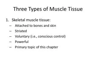
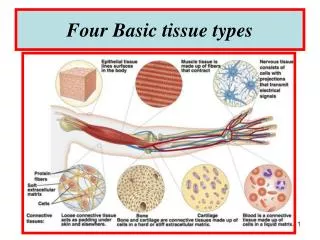
IV. Muscle Tissue • Tissue specialized for contraction • STRAITED VOLUNTARY MUSCLE SKELTAL MUSCLE • STRUCTURE: • Fibers have a band-like appearance created by the protein filaments of actin and myosin • Cells are large, long and multinucleated • Separate cells are hard to see • Is held together by MUSCLE FASCIA • FUNCTION: • Moves bones and other structures VOLUNTARILY when stimulated by nerves • Has the ability to respond to stimuli has “Irritability” • LOCATION: arms, legs, any other muscle that you have control over.
2. NON STRIATED INVOLUNTARY • Structure: • Fibers do not have a band like appearance. • Are not connected to bone • Cells have a single nucleus • Individual cells are visible • Function: • May contract on their own or be stimulated by the involuntary nervous system. • Location: • In the walls of blood vessels • In the iris of the eye • In the gastrointestinal tract
3) STRIATED INVOLUNTARY MUSCLE • CARDIAC MUSCLE Structure: actin and myocin give the cells a striped (striated) appearance. • muscle cells are called CARDIOCYTES and are a branched “network” of interconnected cells. • Cells are multinucleated • Individual cells are hard to see • Are not connected to bone. • Function: • Internal regulator cells “pacemaker cells” causes rhythmic contractions without stimulation that serve to circulate blood through the body • Location: ONLY IN THE HEART
Membranes • At the tissue level (basement membrane) • Form a barrier • Four Types ( are a combination of epithelial and connective tissue) • 1. Mucous • 2. Serous • 3. Cutaneous • 4. Synovial
Mucous Membrane • Structure: • Simple, stratified or transitional epithelium • May contain goblet cells: unicellular exocrine glands or multicellular exocrine glands • Function: • Line cavities that communicate with the exterior environment e.g. digestive, respiratory, reproductive and urinary tract
Serous Membrane • Structure: • Simple epithelium • Loose connective tissue • Very thin and tightly connected to the body wall and organs the cover. • Has parietal and visceral portions: • Parietal: line the outer wall of the internal chamber • Visceral: covers the organs within the body cavity
Serous Membrane (cont) • A transudate or serous fluid covers the parietal and visceral surfaces and minimizes friction between the opposing surfaces. • Function: • To reduce friction between the body wall and internal organs • A) Pleura- lines the pleural cavity and covers lungs • B) Peritoneum- lines the peritoneal cavity and covers the enclosed organs. • C) Pericardium- lines the pericardial cavity and covers the heart. (Pleurisy, Pericarditis, Peritonitis)
Cutaneous Membrane • The skin • Structure: • Stratified squamous epithelium • Underlying connective tissues • Adipose, dense irregular, connective • And accessory organs (glands) • Thick, relatively waterproof and usually dry
Synovial Membranes • Structure: • Loose connective tissue • An incomplete epithelium of squamous or cuboidal cells • There is no basement membrane • Gaps exist between adjacent cells • Function: • Surrounds joints • Regulates the production and composition of SYNOVIAL FLUID which fills the joint cavity and… • Prevents direct contact of bones • Prevents abrasion and damage by impact to the ends of bones • Allows for smooth movement.