Download
1 / 50
500 likes | 648 Views
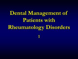
Dental Management of Patients with Rheumatology Disorders. 1. Pathological Classification of Rheumatic Disorders. Rheumatoid arthritis Connective tissue disorder Spondarthritis. Autoimmune Disorder Crystal Arthropathy Infection. Joint Disorder. Inflammatory Disorder.

E N D
Pathological Classification of Rheumatic Disorders Rheumatoid arthritis Connective tissue disorder Spondarthritis Autoimmune Disorder Crystal Arthropathy Infection Joint Disorder Inflammatory Disorder Gouty Arthritis Pseudogout (CPPA) Degenerative Disorder O.A Septic Arthritis
Introduction.. • Is it Arthritis or Arthralgia? • Is it Monoarthritis or Polyarthritis ? • Is it Musculoskeletal emergencies ?
RED FLAG CONDITIONS FRACTURE SEPTIC ARTHRITIS GOUT/PSEUDOGOUT NERVE OR VESSEL PROBLEMS Fever or unexplained weight loss History of carcinoma Immuno-supression Ill health or presence of other medical illness Night pain Progressive pain
Sorting it Out INFLAMMATORY DEGENERATIVE CHRONIC PAIN
What are the Symptoms? Inflammatory Degenerative Chronic Pain Joint Pain Yes Yes No Joint Swelling Yes Yes No Joint Redness Yes No No Morning Stiffness > 1 hour 15-20 minutes > 1 hour Fatigue New and Severe Mild Severe Loss of Function Rapid Slow Rapid Fever Possibly Never Never Weight Loss Possibly Unusual Unusual
Arthralgia.. • Fibromyalgia • Bursitis • Tendinitis • Hypothyroidism • Neuropathic pain • Metabolic bone disease • Depression
Monoarthritis.. • Trauma • Infection: • ± Skin lesion. • Nongonococcal bacterial infections: large joints. • Mycobacterial and fungal infection. • Crystal induced arthritis • Monosodium Urate crystals (MPJ) - Gout • Calcium pyrophosphate dihydrate crystals (knee) - Pseudogout • Systemic Rheumatoid diseases: • Seronegative spodyloarthropathy (Reactive arthritis, psoriatic arthritis, Inflammatory BD..) • RA • Osteoarthritis
Polyarthritis.. • Rheumatoid Arthritis • Systemic lupus Erythrematosus • Viral arthritis • Reiter’s disease • Psoriatic arthritis • Reactive arthritis
Migratory Arthritis.. • Differential diagnosis: • Rheumatic fever • Gonococcemia • Meningococcemia • Viral Arthritis • SLE • Acute Leukemia
Rheumatic Fever.. • Majer Criteria: 1- Carditis 2- Polyarthritis 3- Chorea 4- Erythema Marginatum 5- Subcutaneous nodules • Minor criteria: • 1- Arthralgia 2- Ferver 3- Acute phase reactant (ESR, CRP). • 4- Prolong PR interval 5- Evidence of group A streotococcal infection (AST, Throat culture…)
History.. Age & Sex • <30=SLE, Ankylosis spodylitis, Reactive Arthritis. • 30-50=RA, Systemic sclerosis, Gout. • >50=OA, Pseudogout, PMR • Any Age group=Psoriatic arthritis, Enteropathic arthritis • >Female: SLE, RA, OA, Systemic sclerosis, PMR. • Male=Female: Psoriatic arthritis, Enteropathic arthritis Pseudogout, . • >Male: Gout, Reactive Arthritis, Ankylosis spodylitis,
History.. Symptoms • Site: • Symmetrical= RA, SLE, Systemic sclerosis • Asymmetrical=OA • Large joints= OA • DIP= OA, Psoriatic arthritis • MCP, PIP= RA, SLE • 1st MTP= Gout, OA • Spine= OA, Ankylosis spodylitis, Psoriatic arthritis, Reactive arthritis • Shoulder= PMR
Physical Examination.. • Joint: • Soft tissue swelling, warm, effusion…=Inflammation. • Inflammation signs extended=Septic arthritis, crystalinduced arthritis, fracture. • Passive motion (N), active(↓↓)=Bursitis, Tendinitis, Muscle injury. • Passive motion (↓↓), active(↓↓)=Synovitis
Physical Examination.. • General Examination: • Parotid enlargement, oral ulceration, heart murmurs, pericardial or pleural friction rubs, crackle…=systemic disease. • Fever= Infection, reactive arthritis, RA, SLE, Crystal induced arthritis… • Subcutaneous nodules=RA, RHD, Gout (tophi) • Skin manifestations=Psoriasis, RA, SLE… • Eye disease(keratoconjunctivitis sicca, uveitis. Conjunctivitis, episcleritis…)
Laboratory & Radiology Studies.. • Can be misleading. • Basic: CBC, Urinalysis, U&E, LFT. • Acute phase reactant: ESR, CRP. • Uric acid concentration= Gout • Synovial fluid analysis= infection, crystal induced arthritis, inflammatory.. • Antibody tests: • ANA= SLE • Anti-dsDNA= SLE • Anti-native DNA, anti-Sm= SLE • RF= RA • Anti-CCP antibody=RA • X-ray: • MRI:
Rheumatoid ArthritisAchronic nonsuppurative inflammatory destruction of the joints
Rheumatoid Arthritis.. • Incidence • 1-3% of general population • Genetic predisposition • Female to male ratio 3:1 • Average age of onset of 40 years
History.. • Malaise • Fever • Fatigue • Weight loss • Myalgias • Difficulty performing activities of daily living
Examination.. • Joint affected • swelling • tenderness • warmth • decreased range of motion • Atrophy of the interosseous muscles • deformities
Morning stiffness > 1 hour Arthritis of ≥ 3 joints areas (PIP, MCP, wrist, elbow, knee, ankle, and MTP) Arthritis of hand joints (wrist, MCP, PIP) Symmetric arthritis Rheumatoid nodules RF+ Radiographic changes Erosions Unequivocal periarticular osteopenia ≥ 4 Diagnosis.. ACR Criteria criteria present > 6 wks
Extra-Articular Manifestations.. • Rheumatoid nodule • Cardiovascular • Pulmonary • GI & Renal • Hematological • Skin • Vasculitis • Neurological • Ocular
Ocular • Sicca symptoms • Episcleritis • Scleritis • Scleromalacia Perforance
Head & Neck Manifestations • Rheumatoid Arthritis may involve the TMJ. • 55% Affected 70% with radiographic evidence of TMJ involvement Juvenile form may lead to Retrognathia
Head and Neck Manifestations • Cricoarytenoid joint • Most common cause of cricoarytenoid arthritis • 30% patients hoarse • Exertional dyspnea, ear pain, globus • Hoarseness • Rheumatoid nodules, recurrent nerve involvement • Stridor • local/systemic steroids • Conductive Hearing Loss • Ossicular chain involvement • Sensory Neural Hearing Loss • Unexplained • Assoc. with rheumatoid nodules • Cervical spine • Subluxation
Laboratory .. • Hematologic parameters • Anaemia • Thrombocytosis • ↓ Serum iron & IBC • ↑ Serum globuline • ↑ ALP • ↑ Acute phase reactant ( ESR / CRP ) • Immunological parameters ( RF ) / ANF “50 % ) • Synovial fluid analysis (WBC > 2000/mm3)
Laboratory • Rheumatoid Factor • Ig M Antibody against the Fc fragment of Ig G • Not sensitive • 80% of RA patients • RF+ patients more likely to have • More severe disease • Extraarticular manifestations • Anti-cyclic citrullinated peptide (Anti-CCP ) • Specificity = 90% • Sensitivity = 50-80%
RF is not specific for RA. • Other autoimmune disease • Sjogren’s syndrome , Systemic Lupus • Chronic infection • Hep B/C, SBE, Viral, Parasites, TB • Pulmonary inflammation • Sarcoid, IPF, Silicosis, Asbestosis • Malignancy • Healthy – 4% young; 5-25% > age 60
Radiography • Periarticular osteopenia • Symmetric joint space loss • Marginal erosions • Absence of productive changes • Best films for diagnosis: • Bilateral Hand Arthritis Series • Bilateral Foot Series • Larger joints may not show erosions early due to thicker cartilage.
Treatment • Aggressive Treatment Early! • Physical therapy, daily exercise, splinting, joint protection • Salicylates, NSAIDS, DMARDs , hydroxychloroquine, immunosuppressive agents , Steroids • Cyclosporin-A • Prognosis • 10-15 yrs of disease • 50% fully employed • 10% incapacitated • 10-20% remission • Persistent active cases more than 1 year likely to lead to joint deformities. • Periods of activity cases have better prognosis. • Mortality rate 2.5 times than generalpopulation
Dental Management • Short dental appointments • Assess if Aspirin or NSAIDs are affecting platelet function
Osteoarthritis? Most common form of arthritis Middle-aged to elderly Gradual pain, worse with use F= M up to age 55; after 55 F>M Obesity, history of trauma Cartilage irregularity 10-20% of these symptomatic Only small percentage present for help Joints affected Hands – DIP, PIP, CMC thumb Hips, knees, ankles, great toes Cervical and lumbar spine
Osteoarthritis Mechanical symptoms ( Pain on activity),Stiffness Bony swelling, crepitus DIP (Heberden) PIP (Bouchard) 1st CMCJ, Neck, Lower back, Hips, Knees, 1st MTP Clinical subsets Generalised OA Primary / nodal OA Erosive OA
OsteoarthritisRadiology ( Correlate poorly with symptoms ) Four cardinal features: Joint space narrowing Sclerosis Subchondral cysts Osteophytes
OA Management Pain Relief Simple/compound analgesics, exercises Glucosamine sulphate, patellar taping Topical capsaicin/NSAID; acupuncture Oral NSAIDs – COX2s, gastro-protection Injections – peri-articular, intra-articular Joint Replacement(Referral guidance hip/knee OA ) ? Infection – same day Rapid deterioration/severe disability (2/52 hip, soon – ‘locally agreed’ knee) Symptoms impair QOL – routine Giving way despite Rx– soon (knee only) Acute inflammation (gout, haemarthrosis, pseudogout) – 2/52 (knee only)
Gout? Disease of Monosodium urate crystal deposition in tissues of and around joints Adult men, peaks in ages 40’s to 50’s Urate Overproduction (<10%) vs Under Excretion (90%) Three stages: Asymptomatic hyperuricemia Acute intermittent gout Chronic tophaceous gout Definitive dx by aspiration of fluid
Gout? Onset before 25should raise the question of unusual form of gout , specific enzyme defect A single joint involve in 85-90% of first attack 90% acute attacks in great toe,next in order of frequency are the ankles, heels, knees, wrists, fingers and elbows Acute gouty bursitis-- prepatella, olecranon Chronic Tophi
Septic Arthritis • Septic arthritis is inflammation of a synovial membrane with purulent effusion into the joint capsule, usually due to bacterial infection. • It is an emergency- it can destroy a joint extremely quickly and (v.rarely) lead to sepsis and death • Frequency: • 2-10 cases per 100,000 in the general population. • 30-70 cases per 100,000 in immunosuppressed/ joint prosthesis