Download
1 / 65
650 likes | 666 Views
Mutagen Aneugen Clastogen Carcinogen Teratogen. Mutations. a permanent change in the DNA base-pair or a chromosome Somatic Germ-line Types of mutation Gene mutation Chromosome mutation Genome mutation. Mutations. Spontaneously Induced by chemical mutagens or radiation

E N D
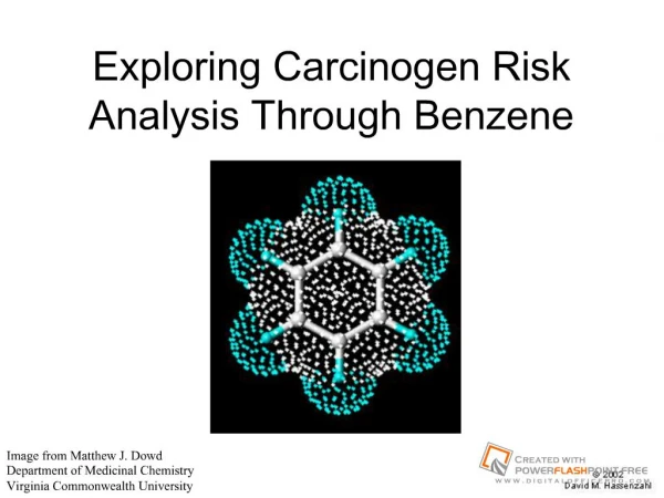
Mutagen • Aneugen • Clastogen • Carcinogen • Teratogen
Mutations • a permanent change in the DNA base-pair or a chromosome • Somatic • Germ-line • Types of mutation • Gene mutation • Chromosome mutation • Genome mutation
Mutations • Spontaneously • Induced by chemical mutagens or radiation • Point mutations: • Silent • Missense • Nonsense • Frameshift mutations
MUTATIONS • Base substitutions • Transition • Pyrimidine changed to another pyrimidine • e.g., C T • Purine changed to another purine • e.g., A G • Transversion • Purines and pyrimidines are interchanged • e.g., A C • More rare than transitions
Mutation: Base Substitution (Point Mutations) P30L in CYP21A2 G Q318X in CYP21A2 G C C • Silent mutation • e.g. L5L in CYP21A2 gene Glu (d) Run-on mutation e.g. Hb Constant Spring TAA>CAA (Stop>Gln)
Mutation Nomenclature c.5162G->A Replacement Original base Base position Deletion: 197delAG Insertion: 2552insT Inversion: 76_83inv p.R197G Original aa del508F in CFTR gene Replacement aa position
Gametogenesis • Gametogenesisgametes • Spermatogenesis • Oogenesis
Spermatogenesis • Occurs in the testes. • Meiosis four cells (sperm)(n=23)
Oogenesis • Occurs in the ovaries and in the oviducts. • After Telophase I and II, the cytoplasm is not equally divided. • Oocyte and polar body
Key Terms • Diploid: Having the full chromosome number (46 in humans). • Haploid: Having half the full chromosome number (23 in humans).
Meiosis Normal Meiosis I Meiosis II
Meiosis I non-disjunction Meiosis I Meiosis II Disomic Nullisomic
Meiosis II non-disjunction Meiosis I Meiosis II Nullisomic Disomic
Non-disjunction • Non-disjunction is the failure of chromosomes (or tetrads) to separate during anaphase I or II in meiosis. • The resulting cells will not have the correct number of chromosomes.
Non-disjunction • If it happens in meiosis I, all the resulting cells will be affected. If it happens in meiosis II, only half will be affected.
Karyotype Indications • advanced maternal age - trisomy risk • chromosomal syndromes (eg., Down syndrome) • mental retardation, developmental delay • abnormal sexual development • repeated spontaneous abortions • Infertility • Malignancy
Karyotype Indications • advanced maternal age - trisomy risk • chromosomal syndromes (eg., Down syndrome) • mental retardation, developmental delay • abnormal sexual development • repeated spontaneous abortions • Infertility • Malignancy
Arm Region Band Subband 3 2 2 1 2 2 1 p 1 5 1 4 1 3 2 1 1 17 q 1 1 . 2 2 1 1 3 1 2 2 q 3 1 3 2, 3 4 1 2 2 4 Chromosome 17 3 Defining Chromosomal Location • ISCN: International System for Human Cytogenetic Nomenclature • Each area of chromosome given number • Lowest number closest (proximal) to centromere • Highest number at tips (distal) to centromere
Chromosome Abnormalities Structural Deletion & microdeletion Duplication Inversion Translocation Reciprocal Robertsonian Isochromosome Ring chromosome Marker chromosome • Numerical • Aneuploid (one extra or missing chromosome) • Polyploid (multiple sets of chromosomes) • Mosaicism (multiple cell lines with different karyotypes)
Tetrad nonsister chromatids chiasmata: site of crossing over Crossing Over
Pericentric inversion • If cross over occurs within the inverted segment • Normal gametes • Balanced gametes • inverted • Unbalanced gametes • Duplication of proximal end & Deletion of distal end • Deletion of proximal end & Duplication of distal end
two identical arms causes Turner syndrome
Numerical abnormalities 2n=46 3n=69 4n=92
Numerical abnormalities • Aneuploidy • The category of chromosome changes which do not involve whole sets. It is usually the consequence of a failure of a single chromosome (or bivalent) to complete division. • Monosomies • All autosomalmonosomies are lethal in very early embryogenesis. They do not even feature in the table of frequencies which follows because they abort too early even to be recognised as a conception. • Trisomies • Crhomosomes 13, 18 and 21 • Down syndrome, trisomy 21 • The incidence of trisomy 21 rises sharply with increasing maternal age.
Aneuploidy caused by • Non-disjunction • failure of homologous chromosomes to separate in anaphase I • failure of sister chromatids to separate at meiosis II • Anaphase lag • Chromosomal loss via micronucleus formation caused by delayed movement of chromosome/chromatid during anaphase • results in daughter cell deficient of that chromosome or chromatid
Meiosis Normal Meiosis I Meiosis II
Meiosis I non-disjunction Meiosis I Meiosis II Disomic Nullisomic
Meiosis II non-disjunction Meiosis I Meiosis II Nullisomic Disomic
Aneuploidy caused by • Non-disjunction • failure of homologous chromosomes to separate in anaphase I • failure of sister chromatids to separate at meiosis II • Anaphase lag • Chromosomal loss via micronucleus formation caused by delayed movement of chromosome/chromatid during anaphase • results in daughter cell deficient of that chromosome or chromatid
Nonclassic modes of inheritance Trinucleotide repeat expansion (dynamic mutations): an unstable heritable element where the probability of expression of a mutant phenotype is a function of the number of copies of the mutation; CGG expansion in Fragile X syndrome FMR1 Gene
Nonclassic modes of inheritance • Biallelic and Monoallelic Expression • Genomic Imprinting: certain genes are expressed in a parent-of-origin-specific manner.
Nonclassic modes of inheritance • Uniparental Disomy: when a person receives two copies of a chromosome, from one parent and no copies from the other parent; e.g Prader-Willi / Angelman syndromes • MatUPD of 15q11-q13 Prader-Willi • PatUPD of 15q11-q13 Angelman syn.
Nonclassic modes of inheritance Mosaicism: condition in which cells within the same person have a different genetic makeup; e.g. germline mosaicism Mitochondrial inheritance: inherited from the mother (maternally inherited)
Genetic Heterogeneity: mutations at different loci (aa and bb at loci A and B); e.g. Ehlers–Danlos syndrome; Hearing Loss • Sometimes, different mutant alleles at same locus very different clinical presentations (Allelic Disorders) • Example: RET gene (encodes a receptor tyrosine kinase) • Some mutations cause dominantly inherited failure of development of colonic ganglia defective colonic motility and severe chronic constipation (Hirschsprung disease) • Other mutations in same gene dominantly inherited cancer of thyroid and adrenal gland (multiple endocrine neoplasia) • A third group of RET mutations both Hirschsprung disease and multiple endocrine neoplasia in the same individual Mutation 1 or 2 or 3 … Disease 1 or 2 or 3 …