Download
1 / 42
430 likes | 643 Views
Optimizing Headache Management in the ED: A Focus on Subarachnoid Hemorrhage. Scott Silvers, MD, FACEP Assistant Professor Co-Director Primary Stroke Center Department of Emergency Medicine Mayo Clinic College of Medicine Jacksonville, Florida. Objectives.

E N D
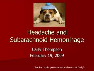
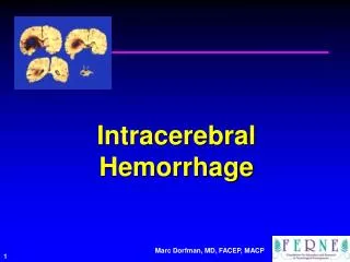
Optimizing Headache Management in the ED: A Focus on Subarachnoid Hemorrhage
Scott Silvers, MD, FACEPAssistantProfessor Co-Director Primary Stroke Center Department of Emergency MedicineMayo Clinic College of MedicineJacksonville, Florida
Objectives • Improve screening of patients for SAH • Learn key points in diagnosis, treatment disposition, documentation • Improve outcome of patients with SAH • Further Emergency Medicine practice as it relates to SAH
Critical Questions • Who is at risk for SAH? • What symptoms suggest SAH? • How can we best diagnose SAH? • Who requires CT? LP? Angiography? • When should an LP be deferred? • When is “traumatic tap” the likely diagnosis? • When does symptom resolution suggest a benign headache etiology?
ACEP Policy: Acute Headache • Does a response to therapy predict the etiology of an acute headache? • Level C: • Pain response to therapy should not be used as the sole diagnostic criteria in determining the underlying etiology of an acute headache. Ann Emerg Med, Jan 2002; 39:108-122
ACEP Policy: Acute Headache • In which adults with a headache can an LP be safely performed without neuroimaging? • Level C: Those pts without signs of increased intracranial pressure (ICP) • Papilledema, absent venous pulses • Altered mental status • Focal neurologic deficits Ann Emerg Med, Jan 2002; 39:108-122
ACEP Policy: Acute Headache • Which patients with an acute headache require neuroimaging? • Level B: • Headache and focal neurologic deficit • Headache of sudden, rapid onset (e.g SAH) • HIV and new headache • Level C: • > 50 years old, new or different headache Ann Emerg Med, Jan 2002; 39:108-122
ACEP Policy: Acute Headache • Do patients with “thunderclap” headache need an angiogram after a negative CT and LP? • Level C: • No, outpatient follow-up if: Negative CT, normal opening pressure, and “negative” CSF analysis Ann Emerg Med, Jan 2002; 39:108-122
Sentinel Headache in SAH • Incidence: ED HA patients = 1/1000 • Present in 10-43% of SAH patients • Typically occurs 2 weeks prior to SAH • Unusual, severe, abrupt, thunderclap • Xanthochromia after first 12 hours Neurol Sci (2004) 25:S 215-217
Sentinel Headache: Symptoms • 77% Nausea/Vomiting • 74% Severe, sudden onset • 64% Focal neuro deficit • 53% Syncope • 33% Stiff neck Neurol Sci (2004) 25:S 215-217
“Worst Headache of My Life” • N= 107 patients “worst headache” • 20 pts with SAH (19.5%) • 18 of 20 diagnosed by CT (90%) • Two diagnosed: + LP after - CT • NPV of CT = 87/89 = 98% (2% would have SAH) Ann Emer Med, Sept 1998; 32: 297-304
“Worst Headache” LP Results • Positive LP, Negative CT (n=2) • Tube 1 RBCs: 163,000 median • Tube 4 RBCs: 221,000 median • Negative LP, Negative CT (N = 77) • Tube 1 RBCs: 19 median • Tube 4 RBCs: 0 median Ann Emer Med, Sept 1998; 32: 297-304
Fifth Generation CT and SAH • 2002 Retrospective study • N = 177 with possible SAH • All pts had both CT and LP • “Fifth generation” CT scanner • “Negative LP” = Tube 1 <400 RBCs and 10-fold drop by tube 4 JEM, 2005; 29: 23-27
Fifth Generation CT and SAH • Results: • 6 CT scans positive for SAH • No CT neg pts had a positive LP • Conclusion: • 5th gen CT detects SAH accurately • 100% sensitivity (61-100%) • 99.4% specificity (97-100%) JEM, 2005; 29: 23-27
SAH: The Evaluation • Evaluate ABCs, altered mental status
SAH: The Evaluation • Evaluate ABCs, altered mental status • Know SAH risk factors: • Hypertension, DM, prior aneurysm/SAH • Thunderclap headache • Maximum severity in minutes • Focal neurological deficit
Non-contrast CT Head • Inform radiologist to rule out SAH • CT should be performed with sufficiently thin cuts (3 – 5 mm cuts) • Unlikely to miss SAH on CT if performed and interpreted well
SAH: The Evaluation • How do we evaluate a CT for SAH?
SAH: CT Interpretation • CT evaluation for subarachnoid blood • 1) Inter-hemispheric fissure • 2) Inferior frontal sulci • 3) Third ventricle • 4) Ambient cistern • 5) Sylvian fissure
Inter-hemispheric fissure Sylvian fissure Cistern blood
CT Interpretation: Elevated ICP • CT findings that exclude elevated ICP • Normal cisterns • No obliteration of cistern space • No edema, mass effect, or midline shift • No hydrocephalus
Symptom Resolution • Can headache resolution be used to exclude SAH? • Brings to mind another question…. In a patient who presents to the ED with a headache, can you rule out SAH by clinical evaluation alone?
Symptom Resolution Consider headaches likely benign if: • Low risk SAH patient • No focal neurological findings • Complete symptom resolution with meds that effectively treat migraine and muscle- tension headache (i.e. non-narcotic)
Lumbar Puncture Need Which patients should have a lumbar puncture?
Lumbar Puncture Indications • Moderate to high risk SAH patients following negative CT • Severe, abrupt, thunderclap headache • Focal neurological findings • Unknown CT protocol / interpretive quality • Minimal symptom resolution with meds that effectively treat migraine and muscle- tension headache
Deferred Lumbar Puncture • Is it sometimes reasonable to not perform a lumbar puncture on patients suspected of SAH?
Deferred Lumbar Puncture • Positive CT • Evidence of elevated ICP, edema, mass effect, midline shift, ICH, hydrocephalus • Technically difficult procedure • Critically ill or unstable patient • Coagulopathy
Measuring Opening Pressure • Is it necessary to measure opening pressure when performing an LP?
Measuring Opening Pressure • Variable practice…. • Measure if CSF flowing rapidly • Consider measuring with every LP
SAH: The Evaluation • How should we interpret CSF results?
Interpreting CSF: RBCs • Likely SAH with: • 10,000-100,000 RBCs or greater • No clearing of RBCs in tube 4 • Consider possible SAH with: • Intermediate RBC count (1,000 – 10,000) • Little RBC clearing by tube 4 • Traumatic tap • 75-90% drop in RBCs from tube 1 to 4
CSF Xanthochromia • Xanthochromia characteristics • Typically > 12 hours from headache onset • Quanitative and qualitative measurements “Read news print test” most often used • Clears after weeks
SAH: The Evaluation • When is angiography indicated?
SAH: Cerebral Angiography • Cerebral angiography indications: • High risk patients with uncertain diagnosis • Interventional radiology available for coiling • Preoperative neurosurgical planning • MRI, MRA, CTA need less well established
SAH: Treatment • How should be treat patients with SAH?
Treating SAH Patients • SAH with increased ICP: • Head of the bed at 45 degrees • Mannitol 20% solution 0.25-1.0g per Kg • Hyperventilation to pCO2 30-35 mmHg, temporizing, only if other measures fail • Ventriculostomy • Consider seizure prophylaxis • Nimodopine (vasoconstriction prophylaxis)
Questions??www.ferne.orgferne@ferne.orgScott Silvers, MD, FACEPsilvers.scott@mayo.edu(904) 296 - 5741 ferne_2005_acep_sa_silvers_BIC_SAH_fshow