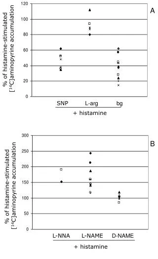
Download
1 / 24
240 likes | 443 Views
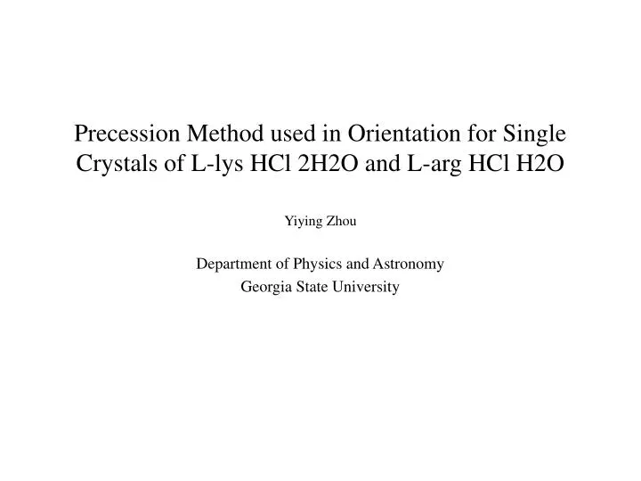
Precession Method used in Orientation for Single Crystals of L-lys HCl 2H2O and L-arg HCl H2O. Yiying Zhou Department of Physics and Astronomy Georgia State University. Outline. What are we study in EPR lab ? Why choose lysine and arginine?

E N D
Precession Method used in Orientation for Single Crystals of L-lys HCl 2H2O and L-arg HCl H2O Yiying Zhou Department of Physics and Astronomy Georgia State University
Outline • What are we study in EPR lab ? • Why choose lysine and arginine? • Precession method used in orienting the crystal of l-lys Hcl 2H2O and l-arg Hcl H2O
1) How to detect the free radicals? • EPR technique is the only analytical technique to measure free radical 2) Why use single crystals? • Single crystals permits taking full advantage of the ESR technique for purposes of radical identification . • The structure information about the molecules and their surroundings can be provided by Crystallography
Molecular Structure of L- arg HCl H2O Unit cell parameters a = 11.044Å b = 8.481Å c = 11.214 Å ß = 91.31 Monoclinic labeling to space group of P21 and there are two molecules in each unit cell.
Molecular Structure of L- lys HCl 2H2O Unit cell parameters: a = 11.044Å b = 8.481Å c = 11.214 Å ß = 91.31 Monoclinic labeling to space group of P21 and there are two molecules in each unit cell.
Distribution of Hydrogen bonds according to the participating amino acids and DNA base or backbone group
It is important to properly locate and assign the axis of crystal • The systems of crystal are anisotropic and thus the magnitude of hyperfine coupling constants, the important magnetic parameters to assign the free radicals, depend strongly on orientation in external magnetic field. • In order to calculate the hyperfine coupling constants (which are generally described by eigenvalues and eigenvectors or tensor) from ENDOR data,it is necessary to take ESR and ENDOR data at lease at two different crystal lattice planes, which means rotate the crystal with two certain different axes and scan the external magnetic field in the plane perpendicular to the rotation axis respectively.
1) Bragg law: N = 2d sin 2) Ewald Construction OP = 1/d410 = (2/ ) sin Principle of Precession Method:
Photos of layer screen and Cirle Circle Layer screen
Sin = (1) t = F (2)
Procession Camera F = 60 mm = 0.7107Å
Diffraction Pattern for single crystal of l-lys Hcl 2H2O (2) (1) (3)
Diffraction pattern for Single crystal of l- arg HCl H2O (1) (2) (3)
Table 1 Parameter of Unit Cell Table 2 Spacing of Diffaction Pattern
l-lys Hcl 2H2O d3 = t3 / (60mm 0.7107Å) (Å-1) d32 (Å-1)2 d32 = (ha*)2 + (kb*)2 + (lc*)2 (Å-1)2 Layer 1 0.1540 0.0237 0.0238 (110)* Layer 2 0.1554 0.0241 0.0238 (110)* Layer3 0.2286 0.0523 0.0523 (021)* l-argHCl H2O d3 = t3 / (60mm 0.7107Å) (Å-1) d32 (Å-1)2 d32 = (ha*)2 + (kb*)2 + (lc*)2 (Å-1)2 Layer1 0.1498 0.0224 0.0222(110)* Layer2 0.1485 0.0220 0.0218(011)* Layer3 0.1488 0.0221 0.0218(011)* Table 3.Comparison of Results of line3
Reference • 1) Luscombe, N, M.; Roman, A.L; Nucleic Acids Research, 2001, 29, • 2860 • 2) Dow, J; Jeansen, L. H.; Acta Cryst., 1970, B26, 1662 • 3) R.R. Bugayong; A. Sequeira; Acta Cryst., 1972, B28, 3214 • 4) A. Ducruix; R. Giege; Crystallization of Nucleic Acids and • Proteins; 1992 Oxford University Press. • 5) Martin J. Buerger; The Procession Method in x-ray crystallograph; • John Wiley and Sons, Inc • 6) John A. Weil; James R. Bolton; Electron Paramagnetic Resonance; • 1994 John Wiley & Sons, Inc.