Download

1 / 37
380 likes | 528 Views
The Lymphatic System. Anatomy & Physiology. The Lymphatic System. Introduction to the Lymphatic System. Two Semi-Independent Parts B. Lymphatic Vessels: transport fluids escaped from the vascular system back to the blood

E N D
The Lymphatic System Anatomy & Physiology
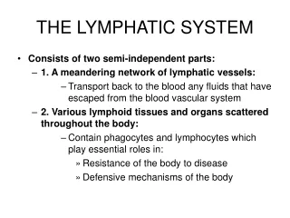
Introduction to the Lymphatic System • Two Semi-Independent Parts B. Lymphatic Vessels: transport fluids escaped from the vascular system back to the blood C. Lymphoid Tissues and Organs: house phagocytic cells, lymphocytes (roles in body defense, disease resistance)
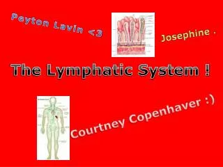
LymphaticVessels A. Special system of drainage vessels which weave between tissue cells and blood capillaries of nearly all tissues B. NOT found in bone, CNS, epidermis, skin, teeth, cartilage, and bone marrow C. Resolve problem of circulatory dynamics of lost fluids and proteins into the interstitial fluid D. Take in cell debris, pathogens, cancer cells E. One way system in which lymph flows only toward the heart F. Blind-ended lymph capillaries with flap-like mini-valves
Lymphatic Vessels…. G. Lacteals: specialized lymphatic capillaries in intestinal mucosa; collect digested fats (chyle = creamy lymph) H. Lymphatic collecting vessels I. Lymphangitis: inflammation of lymphatic vessels; red lines seen through skin that are tender J. Lymphatic trunks: paired lumbar, paired bronchomediastinal, paired subclavian, paired jugular, single intestinal K. Right lymphatic duct: drains lymph from right upper arm, right side of head and thorax L. Thoracic duct: drains lymph from the rest of the body
LymphTransport A. Pumpless B. Milking Action of Skeletal Muscles, Pressure Changes in Thorax, Valves C. Very Low Pressure D. Lymphedema: severe localized edema as result of anything preventing normal return of lymph to blood i.e. tumors, mastectomy
Lymph Fluid Components A. Water B. Plasma Proteins C. Ions D. Lymphoid Cells
Lymphoid Cells A. Lymphocytes 1. T Cells: manage immune response, destroy foreign cells 2. B Cells: produce plasma cells which secrete antibodies B. Macrophages C. Dendritic Cells D. Reticular Cells
Lymph Nodes A. Lymph “Filters” B. Hundreds of Nodes C. Found in Inguinal, Axillary, Cervical Regions of Body, Groin, and Near Gastrointestinal Tract D. Have Phagocytic Macrophages Which Engulf and Destroy Bacteria, Cancer Cells, and Other Foreign Materials
Lymph Node Structure A. Most Are Kidney Shaped and Less Than 2.5 cm in Length B. Divided into Compartments C. B Lymphocytes - Plasma Cells - Release Antibodies D. T Lymphocytes - Circulate - Surveillance Role
Lymphoid Organs A. Spleen 1. Located in left side of abdominal cavity 2. Largest lymphatic organ 3. Lymphocyte proliferation, immune surveillance and response 4. Extract aged, defective blood cells and platelets 5. Removes debris, foreign matter, bacteria, viruses, toxins 6. Stores breakdown products of RBCs for reuse 7. Site of RBC production in fetus 8. Stores blood platelets 9. Splenectomy: surgical removal of spleen; macrophages in liver and bone marrow take of functions of spleen
Lymphoid Organs B. Thymus 1. Found in lower neck region and extends into thorax 2. Causes T Cells to become immunocompetent by secretion of thymosin and thymopoietin 3. Size increases through adolescence; decreases in adulthood; by old age it atrophies and is completely replaced by fatty tissue
Lymphoid Organs Tonsils 1. Ring of lymphatic tissue around the entrance to the pharynx 2. Palatine, lingual, pharyngeal (adenoids) 3. Have invaginations called crypts that trap bacteria and particulate matter 4. “Invite” infection which creates wide variety of immune cells with “memory” for trapped pathogens 5. Lymphocytes remove pathogens, foreign matter
Lymphoid Organs Peyer’sPatches: 1. Lymph nodules found in distal portion of the ileum of the small intestine 2. Also concentrated in wall of appendix 3. Similar to tonsils 4. Destroy bacteria 5. Generate “memory” lymphocytes for long-term immunity Mucosa-Associated Lymphatic Tissue (MALT): • Peyer’spatches, appendix nodules, tonsils, nodules in bronchi • MALT protects the digestive and respiratory systems from foreign matter
Introduction to Immunity A. Immune System: functional system NOT an organ system; recognizes specific foreign substances and acts to immobilize, neutralize, or destroy them B. Immunity 1. Direct: cell attack 2. Indirect: chemicals and antibodies 3. Together result in highly specific resistance to disease
Introduction to Immunity C. Nonspecific (Innate) Defense System 1. Immediate response 2. Two “barricades” 3. First line of defense: external body membranes i.e intact skin and mucosa 4. Second line of defense: called into action by chemical signals when external defenses penetrated a. Uses antimicrobial proteins, phagocytes, other cells b. Hallmark and most important mechanism = inflammation
First Line of Defense A. Intact Skin/Epidermis: forms mechanical barrier that prevents entry of pathogens and other harmful substances into the body 1. Acid Mantle: skin secretions (perspiration, sebum) make epidermal surface acidic which inhibits bacterial growth; sebum contains bactericidal chemicals 2. Keratin: provides resistance against acids, alkalis, and bacterial enzymes
First Line of Defense Intact Mucous Membranes: form mechanical barrier that prevents entry of pathogens 1. Mucus: traps microorganisms in respiratory and digestive tracts 2. Nasal hairs: filter and trap microorganisms in nasal passages 3. Cilia: propel debris-laden mucus away from lower respiratory tract 4. Gastric juice: contains concentrated HCl and protein digesting enzymes that destroy pathogens in stomach 5. Acid mantle of vagina: inhibits growth of bacteria and fungi in female reproductive tract 6. Lacrimal secretions (tears)/saliva: continuously lubricate and cleanse eyes (tears) and oral cavity (saliva); contain lysozyme (enzyme that destroys microorganisms) 7. Urine: normally acid pH inhibits bacterial growth; cleanses lower urinary tract as it flushes from the body
Second Line of Defense Nonspecific Cellular and Chemical Defenses A. Phagocytes: engulf and destroy pathogens that breach surface membrane barriers; macrophages also contribute to immune response B. Natural Killer Cells: promote cell lysis by direct cell attack against virusinfected or cancerous body cells; do not depend on specific antigen recognition
Cont….. Inflammatory Response: prevents spread of injurious agents to adjacent tissues, disposes of pathogens and dead tissue cells, promotes tissue repair; chemical mediators released attract phagocytes (and immunocompetent cells) to the area 1. Inflammatory chemicals: a. Histamine: from granules of basophils; released in response to mechanical injury, presence of certain microbes, and chemicals released by neutrophils; promotes vasodilationof local arterioles, increases permeability of local capillaries permits exudates formation b. Kinins (bradykinin, etc.): peptide formed from plasma protein found in plasma, urine, saliva, lysosomes of some neutrophilsand other cells c. Prostaglandins: fatty acid molecule found in all cell membranes; generated by lysosomal enzymes of neutrophilsand other cells; sensitize blood vessels to effects of other inflammatory mediators
Cont… 2. Four cardinal signs: redness, heat, swelling, pain (5th might be impairment of function) 3. Hyperemia: congestion with blood 4. Exudate: fluid containing proteins such as clotting factors and antibodies 5. Pus: mixture of dead or dying neutrophils, broken down tissue cells, and living and dead pathogens 6. Abscess: inflammatory mechanism fails to clear area of debris; sac of pus walled off by collagen fibers; surgical draining often needed before healing occurs
Cont… D. Antimicrobial Proteins 1. Interferons: proteins released by virus-infected cells that protect uninfected tissue cells from viral takeover; mobilizes the immune system 2. Complement: lyses microorganisms, enhancesphagocytosis, intensifies inflammatory and immune responses E. Fever: systemic response initiated by pyrogens; high body temperature inhibits microbial multiplication and enhances body repair processes
Third Line of Defense: Specific Body Defenses, Immune Response A. Functional system that recognizes foreign molecules (antigens) and acts to immobilize, neutralize, or destroy them B. Antigen Specific: recognizes and is directed against particular pathogens or foreign substances (antigens that incite the immune response) C. Systemic: immunity is not restricted to the initial infection site D. Has Memory: it recognizes and mounts an enhanced attack on previously encountered pathogens
Cells of the Immune System A. B lymphocytes (B cells) 1. Oversee humoral immunity B. T lymphocytes (T cells) 1. Non-antibody producing lymphocytes that constitute cell mediated immunity 2. Extremely mobile, circulate throughout body C. Macrophages 1. Engulf foreign particles and present portions of these antigens on their own surfaces for recognition by lymphocytes 2. Remain in lymph organs
Antigens A. Substances that are capable of mobilizing the immune system and provoking an immune response B. Also, have self-antigens which our bodies recognize and do NOT attack but other bodies would recognize and attack (basis of transplant rejection) C. Immunocompetent: able to recognize a specific antigen by binding to it Immunological Memory A. Primary Immune Response 1. Occurs on first exposure to particular antigen 2. Lag period of 3 – 6 days (time required for the few B cells specific for that antigen to proliferate and for the offspring to differentiate into plasma cells) B. Secondary Immune Response 1. Re-exposure to particular antigen 2. Faster, more prolonged response, more effective 3. Sensitized “memory” cells on alert
Humoral Immunity: Antibody Mediated Immunity (Antibodies in Fluids) A. Antibodies: Immunogloblins - 5 Classes 1. Functions: a. Neutralization: masks dangerous parts of bacterial exotoxins, viruses b. Agglutination: mismatched blood c. Precipitation 2. IgM: first antibody class that is released to blood by plasma cells 3. IgG: most abundant antibody in plasma and only one to cross placenta 4. IgA: found primarily in mucus and other secretions that bather body surfaces; major role in preventing entry of pathogens into body 5. IgD: always bound to B cells; B cell receptor 6. IgE: almost never in blood; “troublemaker” antibodies associated with allergies 7. Hybridomas: fusion of B cells with tumor cells to make monoclonal antibodies; have desirable traits of both parent cells
Humoral Immunity: Antibody Mediated Immunity (Antibodies in Fluids) B. Active Humoral Immunity 1. Naturally acquired during bacterial and viral infections 2. Artificially acquired when person receives vaccines (sometimes booster shots required) - attenuated pathogens usually used C. Passive Humoral Immunity 1. Antibodies harvested NOT made by own plasma cells 2. Acquired by fetus from mother 3. Infusions of immune serum i.e. gamma globulin shots after exposure to hepatitis, antivenoms/antitoxins 4. Donated antibodies provide immediate protection that is short lived (2 – 3 weeks)
Cell Mediated Immune Response A. Lymphocytes B. Causes rejection of grafts or foreign organ transplants unless patient is immunosuppressed C. Infections are the major complications of transplants D. Major histocompatibility complex (MHC): cell surface proteins that mark every body cell as self E. T Cells: cytotoxic T cells (CD8), helper T cells (CD4), and suppressor T-cells (CD8)
Organ Transplants A. Autograft: tissue grafts transplanted from one body site to another in the same person B. Isograft: donated by genetically identical individuals i.e. identical twins C. Allografts: transplanted from individuals that are not genetically identical but belong to the same species D. Xenografts: taken from another animal species i.e. transplanting a baboon heart into a human E. Require immunosuppressive therapy
Homeostatic Imbalances A. Immunodeficiencies 1. AIDS: acquired immune deficiency syndrome; destroys helper T-cells, resulting in depression of cell mediated immunity 2. Congenital thymicaplasia (DiGeorge’s syndrome): thymus fails to develop 3. SCID: severe combined immunodeficiency disease; depletion of both B and T cells B. Hypersensitivities (Allergies) 1. Immediate: i.e. allergy induced asthma 2. Anaphylaxis: life threatening 3. Subacute 4. Delayed: i.e. skin contact with poison ivy, cosmetics, etc.
Cont…. C. Autoimmune Diseases 1. Autoimmune thrombocytopenia: platelets destroyed 2. Multiple sclerosis (MS): destroys white matter of brain and spinal cord; possibly triggered by viral infection; emyelinating disease 3. Myasthenia gravis: impairs communication between nerves and skeletal muscle 4. Graves disease: prompts thyroid gland to produce excessive amount of thyroxine
Cont…. 5. Type 1 (juvenile) diabetes mellitus: possibly triggered by viral infection; destroys pancreatic beta cells - deficit of insulin and inability to use carbohydrates 6. Systemic lupus erythromatosus (SLE); systemic disease that particularly affects the kidneys, lungs, and skin 7. Glomerulonephritis: severe impairment of renal function 8. Rheumatoid arthritis (RA): systematically destroys joints
Testing Time • Make sure to study all your notes for your exam!!