Download
1 / 47
470 likes | 605 Views
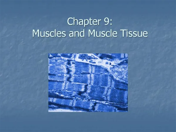
Muscles and Muscle Tissue. Back to aponeurosis. Types of Muscle Tissue. Skeletal muscle is associated with the bony skeleton, and consists of large cells that bear striations and are controlled voluntarily.

E N D
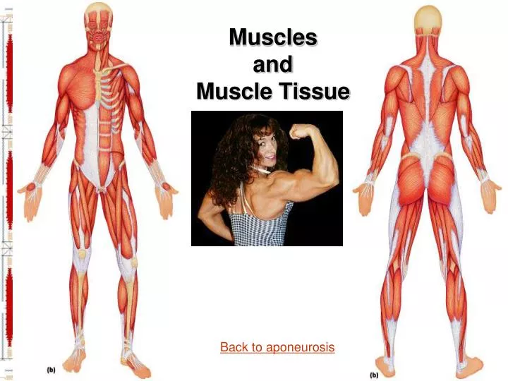
Muscles and Muscle Tissue Back to aponeurosis
Types of Muscle Tissue • Skeletal muscle is associated with the bony skeleton, and consists of large cells that bear striations and are controlled voluntarily. • Cardiac muscle occurs only in the heart, and consists of small cells that are striated and under involuntary control. • Smooth muscleis found in the walls of hollow organs, and consists of small elongated cells that are not striated and are under involuntary control.
Muscle Functions • Movement - bones, pumps. • Muscles aid in maintaining posture. • Muscles stabilize joints by exerting tension around the joint. • Muscles generate heat as a function of their cellular metabolic processes. • Functional Characteristics of Muscle Tissue • Excitability, or irritability. • Contractility. • Extensibility. • Elasticity.
Gross Anatomy of a Muscle Organ • Origin, belly and insertion • The deep fascia connects to bones via tendons & aponeuroses • Made up of thousands of muscle fibers bundled in connective tissue coverings which contains many blood vessels and a motor nerve ending for each muscle fiber • Connective Tissue Wrappings (deep fascia) • Deep fasciasurrounds and penetrates muscle • Epimysium surrounds the muscle organ • Fascia surrounds groups of muscles forming compartments • Perimysium surrounds the fascicles • Endomysium surrounds each muscle fiber • The deep fascia is interconnected to the subcutaneous fascia and the subserous fascia
Skeletal Muscle • Muscles attach: • Directly – epimysium of the muscle is fused to the periosteum of a bone • Indirectly – connective tissue wrappings extend beyond the muscle as a tendon or aponeurosis Return
Muscle Classification: Functional Groups • Skeletal muscles work together or in opposition • Muscles only pull (never push) • As muscles shorten, the insertion generally moves toward the origin • Whatever a muscle (or group of muscles) does, another muscle (or group) “undoes” • Prime movers– provide the major force for producing a specific movement • Antagonists – oppose or reverse a particular movement • Synergists -add force to a movement or reduce undesirable or unnecessary movement • Fixators – synergists that immobilize a bone or muscle’s origin
Arrangement of Fascicles • Parallel – fascicles run parallel to the long axis of the muscle • Fusiform – spindle-shaped muscles • Pennate – short fascicles that attach obliquely to a central tendon running the length of the muscle • Convergent – fascicles converge from a broad origin to a single tendon insertion • Circular – fascicles are arranged in concentric rings
Naming Skeletal Muscles • Location of muscle – bone or body region associated with the muscle • Shape of muscle – e.g., the deltoid muscle (deltoid = triangle) • Relative size – e.g., maximus (largest), minimus (smallest), longus (long) • Direction of fibers – e.g., rectus (fibers run straight), transversus, and oblique (fibers run at angles to an imaginary defined axis) • Number of origins – e.g., biceps (two origins) and triceps (three origins) • Location of attachments – named according to point of origin or insertion • Action – e.g., flexor or extensor, as in the names of muscles that flex or extend, respectively
Microscopic Anatomy of a Skeletal Muscle Fiber • Myofibrils - 80% of the cell • Surrounded by sarcoplasmic reticulum (stores Ca++) • contains transverse tubuleswhich surface at the sarcolemma • Made up of myofilaments • actin -thin • myosin – thick • Each fiber is a long, cylindrical cell with multiple nuclei just beneath the sarcolemma (plasma membrane - supported by dystrophin) • Sarcoplasma - specialties • Glycogen • Myoglobin • mitochondria
Sarcomere -smallest contractile unit • Striations- caused by arrangement of myosin and actin • I band- light • A band - dark • Z line to Z line (one sarcomere) • Molecular composition of myofilaments • myosin • cross bridges (heads contain ATPases) • elongated tail proteins • actin -double stranded helix • Troponin • Tropomyosin • Titin - function • ReviewMatching
Contraction of a Muscle Fiber • Sliding filament model • Which filament moves? • In what direction? 8.
CONTRACTION OF A MUSCLE FIBER • Role of Ca++ in contraction • How is covered up during relaxation? • Where is the binding site for the cross bridge? • How is it exposed for contraction? • Excitation Contraction Coupling • Binding sites exposed (Ca++ present) • Crossbridges form when myosin heads attach using ATP energy • Myosin crossbridges bends pulling on actin using energy from ATP • Crossbridges detach when ATPase hydrolyses new ATP • Ca++ are pumped back into ___________ __________ sacroplasmic reticulum
CONTRACTION REGULATION (neuromuscular junction) 1. At the axon ending a nerve impulse causes the release of ____________ from the synaptic _________. acetylcholine vesicles (Acetylcholine is a ______________) neurotransmitter 2. Acetylcholine crosses the ______ _____ and binds to ___ _________ of the _____ ___ ____ on the sarcolemma. synaptic cleft ACh receptors motor end plate Na+ 3. This opens _____ channels and ____ enter the muscle cell. Na+ 4. Na+ enters the cell and the membrane becomes __________ generating an ______ _________ depolarized action potential 5. The __- _______ carries the action potentialthrough out the cell causing the release of ______from the _________ ___________. T - tubules Ca++ sarcoplasmic reticulum 6. Which then stimulates the _______ _______ action. Sliding filament
Sliding filament model ofcontraction. Myosin heads pull on actin actin moves towards middle of sarcomere How is this controlled? Myosin must attach to actin. Achieved by an increase in Ca+2 in the sarcoplasm
a-The SR & cisternae release Ca+2 into the sarcoplasm b- Ca+2 binds to Troponin C c- The Ca+2 activated Troponin undergoes a shape change that moves tropomyosin away from the myosin- binding site on the actin.
The contraction cycle now begins by: • Myosin head splits ATP & becomes energized 2) Mysosinhead attaches to the myosin binding site on actin (since Ca+2 is present) 3) Myosin head releases the ADP & P, pulls the thin filament toward the center. (power stroke) 4) As the myosin heads bind the ATP, the crossbridges (heads) detach.
Each of the 400-600 myosin heads in one thick filament attaches & detaches about 5 times per second. Not all the crossbridges are bound at the same time.
Resting Membrane Potential • Why does the nerve impulse cross the synapse? • Outside of the cell membrane the electrical charge is positive • Inside the cell the membrane has a negative charge. + • Initially, this is a local electrical event called end plate potential(synapse) • Later, it ignites an action potential that spreads in all directions across the sarcolemma and down the T-Tubules - + + + - + - - - - • The predominant extracellular ion is Na+ • The predominant intracellular ion is K+ • The sarcolemma is relatively impermeable to both ions • All the above facts establish a resting membrane potential
CONTRACTION REGULATION cont. (neuromuscular junction) • Meanwhile back at the junction............................ • The membraneis ___________to accept another stimulus (refractory period) • ____ leaves the cell for a quick repolarization. repolarized K+ • Cholinesterase decomposes ____and removes it from _____________ ACh ACh receptor • Na+-K+ pumpreturnsions to _______ membrane potential conditions resting • In this process where are the 3 places ATP is necessary? • 1. To move myosin crossbridges • 2. To return Ca++ to sacroplasmic reticulum • 3. For the Na+K+ pump to return muscle membrane to resting potential. Review Matching
Motor Unit: Nerve-Muscle Functional Unit • A motor unit is a motor neuron and all the muscle fibers it supplies • The number of muscle fibers per motor unit can vary from four to several hundred • Muscle fibers from a motor unit are spread throughout the muscle; therefore, contraction of a single motor unit causes weak contraction of the entire muscle • Muscles that control fine movements (fingers, eyes) have small motor units • Large weight-bearing muscles (thighs, hips) have large motor units
Motor unit recruitment: not all the MU of a muscle are simultaneously recruited. As force is required for a task #MU are recruited (activated).
Two ways that we produce graded movements-not jerky. 1) MU recruitment 2) frequency of stimulation (APs of nerve & thus muscle cell)
Frequency of stimulation Muscle twitch: response of a MU to a single AP of its motor neuron Wave summation: if another AP occurs before the muscle has had time to fully relax force of contraction
Unfused tetanus: stimulate at a rate of 20-30 x/sec, partial relaxation & force of contraction Fused tetanus: stimulation rate of 80-100x/sec, no relaxation between twitches force of contraction All of the above can be at least partially due to Ca+2 build up in sarcoplasm (hasn’t gotten a chance to get pumped back into SR)
Muscle Twitch • A muscle twitch is the response of a muscle to a single, brief threshold stimulus • Latent period – first few milliseconds after stimulation when excitation-contraction coupling is taking place • Period of contraction – cross bridges actively form and the muscle shortens • Period of relaxation – Ca2+ is reabsorbed into the SR, and muscle tension goes to zero • The three phases of a muscle twitch are:
Graded Muscle Responses • Graded muscle responses are: • Variations in the degree of muscle contraction • Required for proper control of skeletal movement • Responses are graded by: • Changing the frequency of stimulation • More rapidly delivered stimuli result in incomplete tetanus • If stimuli are given quickly enough, complete tetanus results • Changing the strength of the stimulus • Threshold stimulus – the stimulus strength at which the first observable muscle contraction occurs • Beyond threshold, muscle contracts more vigorously as stimulus strength is increased • Force of contraction is precisely controlled by multiple motor unit summation • This phenomenon, called recruitment, brings more and more muscle fibers into play • Starting length of the muscle - optimal length-tension relationship • The relative size of the muscle – the bulkier the muscle, the greater its strength
Contraction of Skeletal Muscle (Organ Level) • The two types of muscle contractions are: • Isometric contraction –Tension increases to the muscle’s capacity, but the muscle neither shortens nor lengthens • Maintain posture • Isotonic contraction – the muscle changes in length (decreasing the angle of the joint) and moves the load • Concentric contractions – the muscle shortens and does work • Eccentric contractions – the muscle contracts as it lengthens • as in setting down a load without dropping it
Muscle Tone & Treppe • Muscle tone: • Is the constant, slightly contracted state of all muscles, which does not produce active movements • Keeps the muscles firm, healthy, and ready to respond to stimulus • Lack of nerve stimulation causes muscles to become flaccid and atrophy. • Spinal reflexes account for muscle tone by: • Activating one motor unit and then another • Responding to activation of stretch receptors in muscles and tendons • Treppe: The Staircase Effect -increased contraction in response to multiple stimuli of the same strength • Contractions increase because: • There is increasing availability of Ca2+ in the sarcoplasm • Muscle enzyme systems become more efficient because heat is increased as muscle contracts
Muscle Metabolism: Energy for Contraction • ATP is the only source used directly for contractile activity • As soon as available stores of ATP are hydrolyzed (4-6 seconds), they are regenerated by: • The interaction of ADP with creatine phosphate (CP), no oxygen required • Anaerobic glycolysis – lactic acid formation, no oxygen required • Aerobic respiration – oxygen required ATP --->ADP + P1 • Heat Production • Only 40% of the energy released in muscle activity is useful as work • The remaining 60% is given off as heat • Dangerous heat levels are prevented by radiation of heat from the skin and sweating
3 Sources of ATP in muscle: 1) creatine phosphate (3 - 6x more plentiful than ATP, only found in muscle) donates a phosphate to ADP. • ATP & creatine phosphate provide energy for 15 sec of maximal activity.
2) Anaerobic (no O2) glycolysis. Glucose (from blood or glycogen breakdown in muscle) is converted to pyruvic acid (get 2 ATP molecules) and then to lactic acid. • Provides energy for 30-40 sec of maximal activity • Both 1 & 2 occur in cytoplasm.
Muscle Metabolism: Anaerobic Glycolysis • When muscle contractile activity reaches 70% of maximum: • Bulging muscles compress blood vessels • Oxygen delivery is impaired • Pyruvic acid is converted into lactic acid • The lactic acid: • Diffuses into the bloodstream • Is picked up and used as fuel by the liver, kidneys, and heart • Is converted back into pyruvic acid by the liver (Cori Cycle)
3) Aerobic(O2 required) Pyruvic acid (from glucose) Amino acids Fatty acids generate ATP (36 per glucose molecule), CO2, H2O, heat. Provides ATP for activities longer than 40 sec Takes place in mitochondria
Muscle Fatigue & Oxygen Debt • Muscle fatigue – the muscle is in a state of physiological inability to contract • Muscle fatigue occurs when: • ATP production fails to keep pace with ATP use • There is a relative deficit of ATP, causing contractures • Lactic acid accumulates in the muscle • Ionic imbalances are present - Na+-K+ pumps cannot restore ionic balances quickly enough • SR is damaged and Ca2+ regulation is disrupted • Intense exercise produces rapid muscle fatigue (with rapid recovery) • Low-intensity exercise produces slow-developing fatigue • Oxygen Debt- the extra amount of O2 needed for a muscle to return to a resting state: • Oxygen reserves must be replenished • Lactic acid must be converted to pyruvic acid – inc. ATP • Glycogen stores must be replaced Cramp
Muscle Fiber Types • Determined by the two following factors: • Contraction rate depends on speed in which ATPases split ATP • Anaerobic (Glycolytic) or Aerobic (oxidative) • Slow-twitch Oxidative Fibers • slow acting myosin ATPases • slow contraction small diameter • always oxidative (aerobic) • resistant to fatigue • red fibers ATP, Mitochondria • capillaries • Posture, endurance, some marathon like • Fast-twitch oxidative fibers • Fast acting myosin ATPases • Fast contraction • moderate resistance to fatigue • pink to red in color • Walking, sprinting • Fast-twitch glycolytic fibers • ATPases fast or slow? dec. ATP • contraction rate? Most force • aerobic or anaerobic? • white fibers (myoglobin?) dec., mitochondria, caps • blood supply? • fatigable? Most jumping, wgt lift
Recruitment order SO, FOG, FG • Most muscles are a mixture of all 3, but percentage varies- genetics and type of exercise. training FG FOG detraining No slow in paralyzed people, Lose fast types as we age & tend to see clustering of types in x- sections.
Smooth Muscle • Composed of spindle-shaped fibers • Lack coarse connective tissue sheaths of skeletal muscle, but have endomysium • Organized into two layers (longitudinal and circular) of closely apposed fibers • Found in walls of hollow organs (except the heart) • When longitudinal layer contracts, the organ dilates & contracts • When the circular layer contracts, the organ elongates – rhythmic contractions • Peristalsis – alternating contractions and relaxations of smooth muscles that mix and squeeze substances through the lumen of hollow organs
Smooth muscle: 2 types 1) Single unit -most common a) walls of hollow organs (except heart)- respiratory, digestive, urinary, reproductive, some blood vessels b) cells arranged in sheets w/ gap junctions between cells- stimulate each other, contract rhythmically
2) multiunit smooth muscle a) large airways, large arteries, irises of eyes b) cells are structurally independent (few gap junctions) so need more innervation
Smooth m cells differ from skeletal m. 1) Cells are smaller,(1000x shorter) tapered 2) No t-tubules, have caveoli: tiny invaginations 3) Actin & myosin are diagonal so don’t see striations
4) No z lines, but intermediate filaments anchored to dense bodies 5) Generate more tension (not limited by sarcomere constraints), but slower to contract/relax 6) > 1 neuron can innervate a cell & > 1 neurotransmitter is possible. No true NMJ- varicosities
7) Ca+2 binds to calmodulin (not troponin) This Ca-calmodulin complex activates an enzyme (kinase) which puts a PO4 on the mysosin head & now it can interact with actin
Innervation of Smooth Muscle • Smooth muscle lacks neuromuscular junctions • Innervating nerves have bulbous swellings called varicosities • Varicosities release neurotransmitters into wide synaptic clefts called diffuse junctions • Whole sheets of smooth muscle exhibit slow, synchronized contraction • They contract in unison, reflecting their electrical coupling with gap junctions • Action potentials are transmitted from cell to cell
Characteristics of Smooth Muscle • Unique characteristics of smooth muscle include: • Smooth muscle tone • Slow, prolonged contractile activity • Low energy requirements • Response to stretch • Smooth muscle exhibits stress-relaxation response (compliance) in which: • Smooth muscle responds to stretch briefly, and adapts to its new length • The new length, however, retains its ability to contract • This enables organs such as the stomach and bladder to temporarily store contents • Smooth muscle has good regenerative ability • This is shown by estrogen’s effect on the uterus • At puberty, estrogen stimulates the synthesis of more smooth muscle, causing the uterus to grow to adult size • During pregnancy, estrogen stimulates uterine growth to accommodate the increasing size of the growing fetus • Autonomic nervous system and endocrine systems are major controls • Neurotransmitters and hormones?
Developmental Aspects • Nearly all muscle tissue develops from specialized mesodermal cells called myoblasts. • Skeletal muscle fibers form through the fusion of several myoblasts, and are actively contracting by week 7 of fetal development. • Myoblasts of cardiac and smooth muscle do not fuse but form gap junctions at a very early stage. • Muscular development in infants is mostly reflexive at birth, and progresses in a head-to-toe and proximal-to-distal direction. • Women have relatively less muscle mass than men due to the effects of the male sex hormone testosterone, which accounts for the difference in strength between the sexes. • Muscular dystrophy is one of the few disorders that muscles experience, and is characterized by atrophy and degeneration of muscle tissue. Enlargement of muscles is due to fat and connective tissue deposit.
References • Muscle Powerpoints • GetBodySmart • Gateway Community College • Muscle Physiology • Lumen • Muscle Models • Marieb Muscle Exercise