Download
1 / 24
270 likes | 340 Views
Cysticercosis and echinococcosis Paul R. Earl Facultad de Ciencias Biológicas Universidad Autónoma de Nuevo León San Nicolás, NL 66451, Mexico.

E N D
Cysticercosis and echinococcosisPaul R. EarlFacultad de Ciencias BiológicasUniversidad Autónoma de Nuevo LeónSan Nicolás, NL 66451, Mexico
Tapeworms(cestodes) are parasites of the intestinal tract whose simple life cycle requires at least one intermediate host. They are long, flat worms that have a scolex or sucker located on the head, armed with hooks or not. The cestodes that cause significant human disease include Taenia saginata of cattle, Taenia solium of pigs, Hymenolepsis nana of mice and others, and Diphyllobothrium latum of fishes. The latter 2 members of the Cestoda are not considered further here.
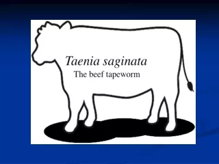
T. saginata, the most common tapeworm in humans, is found wherever raw or undercooked beef is eaten, especially in Africa and the Arab world In certain cultures where raw beef is considered a delicacy, the number of tapeworms harbored may be considered a status symbol. Cattle become infected in areas where raw sewage (aguas negras) drains into pastures. Chemical treatment of the soil does not kill the ova, which may live in the soil for up to 6 months.
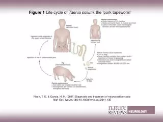
Life cycle. Humans are the definitive host of T. saginata, and infection occurs after ingestion of beef containing viable larvas. The larvas are released with digestion and mature in the upper small intestine. Adult worms may inhabit the intestinal tract for as long as 25 years. The adult worms release eggs that are passed through the feces onto soil and vegetation. Herbivorous animals ingest the eggs, which penetrate the intestinal tract and migrate to the skeletal muscle.
Clinical manifestations. The majority of T. saginata infections are asymptomatic. The most common symptoms are epigastric pain, nausea, vomiting, increased appetite, weight loss, and a spontaneous emerging of proglottid on the anus. Intestinal obstruction is a rare complication.
Diagnosis, treatment, and prevention.The diagnosis is made by finding typical eggs in the host stool. The eggs of T. saginata and T. solium are indistinguishable; however, the two may be differentiated through examination of gravid proglottids pressed between two glass slides. If more than 13 uterine branches are present on each side of the proglottid, the infection is consistent with T. saginata.
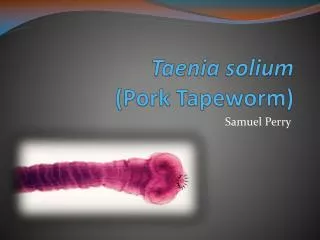
T. solium infection occurs in all areas where raw or partially cooked pork is eaten. Usually, clinical cysticercosis refers to disease caused by T. solium. It is common throughout Mexico, South America and southern Europe. T. solium infection is uncommon in the United States, but of course does occur.
Life cycle. The life cycle of T. solium is remarkably similar to that of T.saginata with the primary difference being the intermediate host, the pig. Adult worms may inhabit the human upper intestinal tract for as long as 25 years. Clinical manifestations.T. solium infection is usually asymptomatic. The most common intestinal symptoms include abdominal pain, nausea, vomiting and diarrhea. One of the most serious complications is cysticercosis, an infection of the larval forms of T. solium, which invade the brain and other organs.
Diagnosis, treatment, and prevention.The diagnosis of T. solium infection is made by the identification of typical eggs in the host’s stool or by the use of adhesive tape placed on the anus. The treatment of T. solium infection is the same as that for T. saginata. Prevention of T. solium infection can be achieved with adequate cooking of all pork and pork products. Cysticerci develop in the brain, very rarely in other organs
The eggs of Taenia solium and T. sagiata look the same.Armed scolex of Taenia solium with rostrellum and hooklets.
Proglottid of Taenia solium Taenia saginata without rostellum, unarmed. Proglottid of Taenia saginata
Cystycercosis.The World Health Organization (WHO) estimates that 50 million people are infected by the cestode teniasis/cysticercosis complex, and that 50,000 of them die each year (0.1 %). Rustic techniques for raising pigs are one of many factors in disease transmission. Many human populations show infection rates of 5-15 % serologically. Often symptomless, cysticercosis is always underreported and thus the prevalence is rarely known. The occurrence of cysts in the brain is the dramatic feature.
In a Mexican study of 10,000 cases of cysticerosis, men were 59.30 % and women 40.70 % of the population having 9,996 Mexicans, the rest foreigners. Sixty percent of cases were in the middle class, 10 % in the upper class and 30% among the poor. About 60 % of cysts were alive and 40 % calcified. Importantly, 13 % were previously treated with praziquantal with many complications and required a second treatment with radioimmune methods.
The symptoms of the anterior cases were: a) 62 % headache, b) 25 % seizures, c) 12 % endocranial hypertension, d) 1% pseudotumor, e) 1 % medullar shock and f) 1 % miscellaney. Organs with cysts were: a) 97.82 % brain, b) 1.00 % muscle, c) 0.90 % subcutaneus tissue, d) 0.02 kidney, e) 0.01 % heart, f) 0.03 % submaxillary salivary glands, g) 0.008 % eye and h) 0.001 liver.
Section of brain by Ana Flisser, Universidad Nacional Autónoma de México, Mexico X-ray plate showing cyst
Echinococcosis.Regarding echinococcosis, Central Europe as western France and eastern Austria with 210 patients, 61.4 % worked as farmers, gardeners, foresters or hunters, 78 % having symptoms. Alveolar echinococcosis was primarily manifested as a liver disease. Of the 559 patients, 190 (34%) were already affected by spread of the parasitic larval tissue. Of 408 (73%) patients alive in 2000, 4.9% were cured. The increasing prevalence of E. multilocularis corresponds with the increase in foxes.
Echinococcosis in humans is a complex of parasitic diseases caused by the larval stages of 4 species of the cestode genus Echinococcus, which are perpetuated in life-cycles, involving domestic and wild carnivores like foxes as definitive hosts and a wide range of mammals (livestock animals, rodents, etc.) as intermediate hosts. Echinococcosis is not only one of the most devastating parasitic diseases of humans, but it is also very difficult and costly to treat, control and prevent.
Cystic echinococcosis, caused by Echinococcusgranulosus and E. multilocurlaris, remains a considerable public health (PH) problem in many countries. These respective diseases are alveolar and cystic hydatidosis. In the countries of Central and South America, human polycystic echinococcosis occurs caused by Echinococcus vogeli and E. oligarthrus.
Juvenile proglottids of Echinococcus granulosus Echinococcus granulosus
Echinococcosis is mainly caused by larvas of 2 species of small tapeworms, Echinococcusgranulosus (cystic echinococcosis) andE. multilocularis (alveolar echinococcosis). E. granulosusoccurs worldwide, and E. multilocularis is found in the NorthernHemisphere. Humans can develop the diseases when they ingest eggsexcreted with the feces of the final hosts (dogs and foxes). E.granulosus larvae then grow as large cysts with internal buddingof brood capsules.
Detection, serology and related.Eggs and proglottids in feces are commonly used for detection. The EchinococcusWestern Blot IgG using a whole larval antigen from E. multiloculariswas evaluated for serodiagnosis and differentiation between 2human parasitic infections of worldwide importance: a) cystic echinococcosisdue to E. granulosus and b) alveolar echinococcosis, dueto E. multilocularis. The assay allowedthe detection of serum immunoglobulin G (IgG) antibodies in 97% of Echinococcus-infected patients. E. granulosus wascorrectly distinguished from E. multilocularis-infectedpatients in 76% of cases.
Education.A propoganda campaign as a warning on handling swine and cattle, and of course raw and undercooked meats can reduce cases, especially if accompanied by a deparasitizing campaign for pigs, and even for school children. For instance, posters of the life cycle on country fence posts might be appliciple. Control just by deparasitizing pigs might be sufficient to truely sanitize the environment. Ignorance of the danger of cysts in the brain is the main problem. Can PH education and deparasitizations reduce the number of operated cases of neurocysticercosis so that they actually pay for themselves?