Download
1 / 75
800 likes | 904 Views
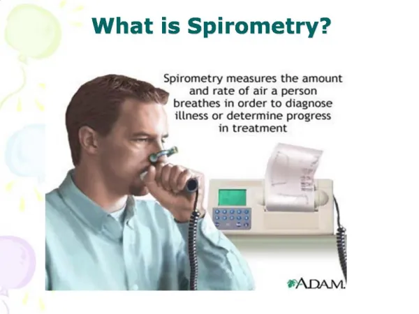
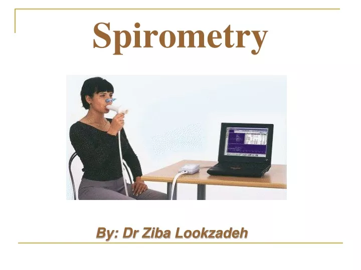
Spirometry. By: Dr Ziba Lookzadeh. Introduction. A non-invasive method for evaluation of pulmonary function Simple, cost-effective, accessible Not for definite diagnosis of disease but help diagnosis along with history, physical examination and other paraclinical diagnostic method.

E N D
Spirometry By:Dr ZibaLookzadeh
Introduction • A non-invasive method for evaluation of pulmonary function • Simple, cost-effective, accessible • Not for definite diagnosis of disease but help diagnosis along with history, physical examination and other paraclinical diagnostic method
Spirometry as a screening method • Primary prevention • Pre-placement and fitness-for-duty examinations • Physical demands of a job (heavy manual labor, fire fighting); • Characteristics of respiratory use (prolonged use of negative-pressure mask under conditions of heavy physical exertion and/or heat stress - not required by OSHA); • Research and monitoring of health status in groups of workers.
Spirometry as a screening method • Secondary prevention • Medical surveillance programs – workers at risk of developing occupationally related respiratory disorders • Baseline and periodic evaluations • Mandated OSHA regulations (asbestos, cadmium, coke oven emissions or cotton dust) • Local mandated medical surveillance program • Component of workplace health promotion program
Spirometry as a screening method • Tertiary prevention • Clinical evaluation of symptomatic individuals • Restrictive • Obstructive • Combined ventilatory defects • Disability under Social Security Administration • Workers’ compensation setting
Contraindications of Spirometry • Uncontrolled hypertension • Suspected presence of active TB other communicable respiratory disease • Thoracic or abdominal surgery within recent 3 wks • MI or unstable angina within recent 6 wks • Respiratory distress • Active hemoptysis • Recent eye/ear surgery or ear drum perforation • Abdominal or thoracic aortic aneurysm
Confounding factors • Common cold (3 days ego) • Severe respiratory infection (3w) • Smoking( 1hr) • Heavy food (1hr) • Bronchodilator use
Complications • Chest pain • Syncope, dizziness • Increased ICP • Paroxysmal coughing • Nosocomial infection • Bronchospasm
Lung volumes • TV :The volume of air inhaled & exhaled at each breath during normal quiet breathing • IRV: The maximum amount of air that can be inhaled after a normal inhalation • ERV: The volume of air that can be forcefully expired following a normal quiet expiration • RV: The volume of air remaining in the lungs after a forceful expiration
Lung capacities • TLC: The total volume of the lungs • VC: The maximum amount of air that can be exhaled after the fullest inspiration possible • IC :The maximum of air that can be inhale after end tidal position • FRC: The amount of air remaining in the lungs after a normal quiet expiration
Spirometry Parameters • Forced Vital Capacity • FVC • Forced Expiratory Volume in One Second • FEV1 • Forced Expiratory Volume in One Second Expressed as a Percentage of the Forced Vital Capacity • FEV1/FVC % • Mean Forced Expiratory Flow during the Middle Half of the Forced Vital Capacity • FEF 25-75%
FVC • Definition: • Defined as the maximal amount of air that can be exhaled forcefully after a maximal inspiration or the most air a person can blow out after taking the deepest possible breath.
FVC - forced vital capacity • defines maximum volume of exchangeable air in lung (vital capacity) • forced expiratory breathing maneuver • requires muscular effort and some patient training • initial (healthy) FVC values approx 4 liters • slowly diminishes with normal aging • significantly reduced FVC suggests damage to lung parenchyma • restrictive lung disease (fibrosis) • loss of functional alveolar tissue (atelectasis) • intra-subject variability factors • age • sex • height • ethnicity
FEV1 • Definition: • The volume of air exhaled during the first second of a forced expiratory maneuver. • normal FEV1 about 3 liters
FEV1/FVC% • Definition: • The value expresses the volume of air the worker exhales in one second as a percent of the total volume of air that is exhaled. • Calculated by using largest valid FEV1 and largest FVC even if they are not from the same tracing. • Find largest valid FEV1 • Find largest valid FVC • Divide FEV1 by FVC • Multiply by 100 to obtain percentage.
FEF25-75% • Definition: • The mean expiratory flow during the middle half of the FVC • More sensitive than FEV1. • Considerably more variability than FVC and FEV1. • ATS recommends only be considered after determining presence and clinical severity of impairment and should not be used to diagnosis disease in individual patients
PEF - Peak Expiratory Flow rate • measures airflow limitations in large (central) airways • PEF measurements recommended for asthma management • spirometry is recommended to help make the diagnosis of asthma • PEF not recommend to evaluate patients for COPD • cannot measure small airway airflow limitations • advantages of PEF tests • measurements within a minute (three short breaths) • uses simple, safe, hand-held devices that typical, costs $20 • disadvantages of PEF tests (compared to spirometry) • insensitive to obstruction of small airways (mild or early obstruction) • PEF is very dependent on patient effort (large intra-subject variability) • mechanical PEF meters are much less accurate than spirometers
The Original Wright Peak Flow Meter - Standard and Low Range versions
The Mini Wright Peak Flow Meter - Standard and Low Range versions (from left to right: Wright scale, EU scale, ATS scale)
FEV1 FVC PEF FEF25-75% V-T Curve F-V loop Most important parameters
Spirometry Performance Steps • Equipment performance criteria • Equipment quality control ( calibration & leak ) • Contraindications & interfering condition • Age, height, race, gender • Selection of appropriate reference value • Patient maneuver • Acceptability criteria • Reproducibility criteria • Selection of best curve and best result • interpretation
Equipment quality control ( flow-type calibration) • جهت انجام بررسي وضعيت کاليبراسيون از سرنگ 3 ليتري مخصوص استفاده مي شود. • از منوي دستگاه “calibration check ”- option را انتخاب کنيد (جهت حذف فاکتور اصلاح BTPS). • براي بررسي وضعيت کاليبراسيون اسپيرومترهاي flow- type بايد کاليبراسيون را باسه سرعت مختلف انجام داد. يکبار 3 ليتر را به مدت يک ثانيه (سریع) و بار دوم 3 ليتر را در مدت 2-3 ثانيه (سرعت متوسط) و بار آخر 3 ليتر را در مدت 6 ثانيه (سرعت آهسته) به دستگاه تزريق کنيد. در هر سه بار باید عدد ثبت شده FVC بين 2.91-3.09 ليتر باشد. در غير اينصورت کاليبراسيون دستگاه اشکال دارد . • توجه: نکات مذکور اصول استاندارد نحوه کنترل کاليبراسيون و نشت مي باشد ولي در عين حال توصيه مي شود از کتابچه راهنماي دستگاه اسپيرومتر خريداري شده، در مورد نحوه کاليبراسيون و نشت، توصيه هاي کارخانه سازنده دستگاه رانيز مطالعه کرده و آن را نيز در نظر بگيريد.
Equipment validation • Calibration: daily if for screening every 4hr
Subject’s position: • Sitting or standing? • Chair with arms & without wheels • Clothing • Chin & neck position • Nose clip • Denture
Good Start of Test • Start of test must be quick and forceful • No excessive hesitation • Best evaluated using the Flow-Volume tracing • No excessive back extrapolated volume
No Coughing • Especially during the first 1 second of the maneuver • Best if no coughing present during maneuver, however: • Some patients cough near the end of each test, if present then document
No Variable Flow Flow rate should be maximal and consistent throughout testing Volume-Time and Flow-Volume tracings should be smooth