Download
1 / 42
420 likes | 426 Views
Introduction to the study of the nervous system Meninges , hemispheres, the lateral ventricles. Dr. Andrea D. Székely Semmelweis University Faculty of Medicine Department of Anatomy, Histology and Embryology Budapest. WHAT IS THIS NEURO „STUFF ” ABOUT?

E N D
Introduction to the study of the nervous system Meninges, hemispheres, the lateral ventricles Dr. Andrea D.Székely Semmelweis University Faculty of Medicine Department of Anatomy, Histology and Embryology Budapest
WHAT IS THIS NEURO „STUFF” ABOUT? NEUROSCIENCE? NEUROBIOLOGY? NEUROANATOMY? (NEUROLOGY?)
Core Text of Neuroanatomy. 3rd edition. By MALCOLM B. CARPENTER. (Pp. xiv +437; numerous illustrations; £18.00.) Baltimore, London and Sydney: Williams& Wilkins. 1985. This is a well produced textbook with excellent illustrations and a lucid text. Unfortunately,despite its title it is far too detailed for most undergraduate neuroanatomy courses in the BritishIsles. In his review of the first edition (J. Anat. 115, 1973) Professor Sinclair complained of thedetailed content. Since that time the content has continued to expand and indeed the currentedition is more than a third larger than the second edition. This would be a useful book to have on the departmental bookshelf as a reference book forhonours students who require an up to date and well documented description of neuroanatomyat a price departments can afford in these times of financial stringency. R. R. STURROCK J. Anatomy, 1986
NEUROSCIENCE Traditionally, neuroscience is a branch of biology. It is an interdisciplinary science that collaborates with other fields such as chemistry, cognitive science, computer science, engineering, linguistics, mathematics, medicine (including neurology), genetics, and allied disciplines including philosophy, physics, and psychology. It also exerts influence on other fields, such as neuroeducation, neuroethics, and neurolaw. wikipedia Ramon y Cajal
NEUROANATOMY It is concerned with the study of the morphology of the nervous system (CNS, PNS) Both macroscopy and microscopy Functional relevance Comparative neuroanatomy (slipping into neuroscience) HISTORYFromthefirst known written record: Egyptian document Edwin Smith Papyrus stretches over thousands of yearsuptonowe.g. FUNCTIONAL MRI STUDIES GiulioCasserio (c.1545-1616) The firstconcise drawing of the circle of Willis ConstanzoVarolio (1543-1575) Namesthepons = bridge(ponsVaroli)
COMPONENTS OF THE NERVOUS SYSTEM CNS AND PNS neurocranium Peripheralnerve plexi („plexuses”) Vertebralcanal
BRAIN IN SITU Dura mater encephali Foramenmagnum Dura mater spinalis Cerebralfalx
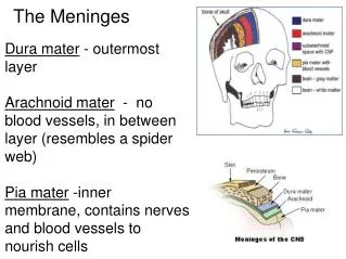
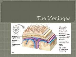
Dura mater MEMBRANES OF THE BRAIN = MENINGES Arachnoid mater Pia mater
MEMBRANES OF THE BRAIN = MENINGES PACHYMENINX (1) The dura mater is a sac that envelops the arachnoid mater and surrounds and supports the large dural sinuses carryingvenous blood. EXTERNAL LAYER - loosely arranged, fibroelastic layer of cells, multipleinterdigitating cell processes, no extracellular collagen, and significant extracellular spaces. The „middle”layer- mostly fibrous. It consists of two layers, wheretheinnerlayer is composed of dense fibrous tissue, withthe inner surface being covered by flattened cells like those present on the surfaces of the pia mater and arachnoid mater. This thin, transparent membrane is composed of fibrous tissue and, like the pia mater, is covered by flat cells also thought to be impermeable to fluid.Thus, itrepresent an effective morphological and physiological meningeal barrier between the cerebrospinal fluid and subarachnoid space and the blood circulation in the dura. It is the meningeal envelope that firmly adheres to the surface of the brain and spinal cord, following all of the brain's contours (the gyri and sulci). It is a very thin membrane, composed of fibrous tissue, covered on its outer surface by a sheet of flat cells thought to be impermeable to fluid.Internallyitcomesincontactwiththe limiting glialmembrane. Dura mater - periosteallayer - meningeallayer Subduralspace (virtualspace) Arachnoid mater Subarachnoidalspace (CSF) Pia mater LEPTOMENINX (2)
DURAL INFOLDINGS • falxcerebri • tentoriumcerebelli • falxcerebelli • diaphragmasellae
DURAL SINUSES & TRIBUTARIES sinus sagittalissuperior - vv. cerebrisuperiores sinus sagittalisinferior sinus rectus - v. cerebrimagnaGaleni sinus occipitalis confluenssinuum sinus transversus - vv. cerebriinferiores sinus sigmoideus sinus petrosussuperior - vv. cerebriinferiores sinus petrosusinferior - v. labyrinthi sinus sphenoparietalis + vv. duraematris sinus cavernosus - vv. cerebriinferiores v. cerebrimedialissuperficialis v. ophtalmicasuperior
DEVELOPMENT OF THE BRAIN Forebrain Midbrain Hindbrain
WITHIN THE CONVEXITY LIES A DEEP CORTICAL PORTION - THE INSULA
MEDULLA - WHITE MATTER FIBRE TRACTS
projection of thebasalgangliaontothebrainsurface and ventricularsystem
CASTING OF THE VENTRICLES AND THECHOROIDAL PLEXUS PARS CENTRALIS ANTERIOR HORN POSTERIOR HORN INFERIOR HORN
WALLS OF THE LATERAL VENTRICLE ANTERIOR HORN Roof: Corpus callosum Lateral wall and floor: Caudate nucleus Medial wall: septum pellucidum PARS CENTRALIS Roof: Corpus callosum. Floor: dorsal surface of thalamus Medial wall and floor: fornix Lateral wall and floor: body and tail of the caudate nucleus INFERIOR HORN POSTERIOR HORN Roof: tail of caudate nucleus, corpus callosum and stria terminalis Floor: hippocampus, collateral eminence, fimbria hippocampi. Lateral wall: tapetum, geniculocalcarine tract, and arcuate fasciculus of the cerebral hemisphere Roof: Corpus callosum Lateral wall: Tapetum (of splenium) Floor: Collateral trigone Medial wall: calcar avis
teniaterminalis teniachoroidea teniafimbriae teniafornicis