Download
1 / 56
560 likes | 623 Views
The Nervous System. Chapter 7. Overview. The nervous system allows the body to detect, interpret and respond to stimuli. A stimulus is any change in the external or internal environment. Functions of the Nervous System. 1. Sensory input

E N D
The Nervous System Chapter 7
Overview • The nervous system allows the body to detect, interpret and respond to stimuli. • A stimulus is any change in the external or internal environment.
Functions of the Nervous System 1. Sensory input • To monitor changes occurring inside and outside the body using sensoryreceptors 2. Integration • To process and interpret sensory input 3. Motor output • To produce a response, if needed, that activates muscles or glands (effectors)
Organization of the Nervous System • Two main divisions: • Central nervous system (CNS) • Brain • Spinal cord • Peripheral nervous system (PNS) • Nerves outside the brain and spinal cord
Nervous Tissue • Nervous tissue is made up of neuroglial(supporting ) cells and neurons. • Neuroglial cells outnumber neurons. • Neuroglial cells do not conduct impulses; neurons do. • Neuroglial cells generally support, insulate and protect delicate neurons. • Neuroglial cells are capable of dividing; neurons are not.
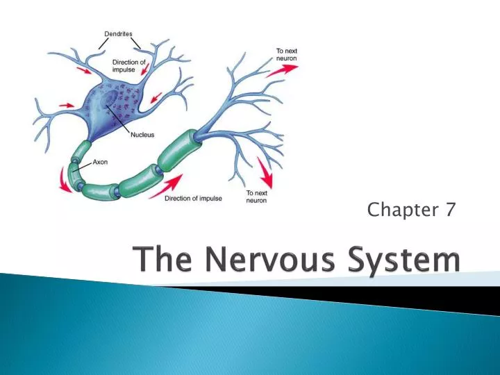
Neurons • Neurons = nerve cells • Specialized to transmit messages (impulses) • Major regions of neurons: • Cell body – nucleus and metabolic center of the cell • Processes – fibers that extend from the cell body • Dendrites– carry impulses towards the cell body • Axon – carries impulse away from the cell body
Neurons • Myelin sheath – fatty material surrounding axons produced by Schwann cells • Insulates axons • Speeds up transmission
Classification of Neurons • Sensory (afferent) neurons • Carry impulses from sensory receptors to the CNS • Motor (efferent) neurons • Carry impulses from the CNS to muscles or glands (effectors) • Interneurons (associative neurons) • Connect sensory and motor neurons
The Nerve Impulse • The plasma membrane at rest is polarized (resting potential) • Fewer positive ions are inside the cell than outside the cell • Active transport by the sodium-potassium pump maintains this polarity
Starting the Nerve Impulse • A stimulus depolarizes the neuron’s membrane • Sodium ions (Na+) rush inside the membrane • This initiates an impulse (action potential)in the neuron
Continuing the Nerve Impulse • The impulse continues to move toward the cell body • Potassium ions rush out of the neuron which repolarizes the membrane • The sodium-potassium pump thenestablishes the original polarity
The Threshold • A minimum stimulus (threshold) is needed to start an impulse • If the threshold is met, a nerve impulse starts, and continues over the entire axon (all or none response) • Impulses travel faster when fibers have a myelin sheath
Neuron to Neuron Communication • Neurons do not touch each other because of a gap called the synapse. • Impulses cross the synapse with the help of chemicals called neurotransmitters. • Neurotransmitters released from the axon terminal diffuse across the synapse • The dendrite of the next neuron has receptors that are stimulated by the neurotransmitter • An action potential (impulse) is started
The Central Nervous System • Brain and spinal cord
Regions of the Brain • Cerebral hemispheres • Diencephalon • Brain stem • Cerebellum
Cerebrum • Cerebrum • Voluntary activities (motor initiation), intelligence, learning, judgment, sensory interpretation • Two hemispheres connected by the corpus callosum • Right hemisphere controls the left side of the body and visa-versa • Deep folds and grooves increase surface area • Gray matter – outer layer of dense nerve cell bodies • White matter – inner layer of myelinated nerve fibers
Lobes of the Cerebrum • Fissures (deep grooves) divide the cerebrum into lobes • Surface lobes of the cerebrum • Frontal lobe • Parietal lobe • Occipital lobe • Temporal lobe
Specialized Areas of the Cerebrum • Primary sensory area (postcentralgyrus)– receives impulses from the body’s sensory receptors • Primary motor area (precentralgyrus)– sends impulses to skeletal muscles
Specialized Area of the Cerebrum • Cerebral areas involved in special senses • Broca’s area – speech • Gustatory area - taste • Visual area - sight • Auditory area - hearing • Olfactory area – smell • Interpretation areas of the cerebrum • Speech/language region • Language comprehension region • General interpretation area
Cerebellum • Two hemispheres with convoluted surfaces • Provides involuntary coordination of body movements • http://www.youtube.com/watch?feature=player_detailpage&v=F7Yw26zHoEw
Diencephalon • Sits on top of the brain stem • Enclosed by the cerebral hemispheres • Made of three parts • Thalamus – sensory relay station • Hypothalamus – controls hunger, thirst, fatigue, anger, temperature, coordinates with endocrine system • Epithalamus – pineal gland and choroid plexus (forms cerebrospinal fluid)
Brain Stem • Attaches to the spinal cord • Controls vital functions – blood pressure, heart rate, breathing, swallowing, vomiting • Parts of the brain stem • Midbrain • Pons • Medulla oblongata
Protection of the Central Nervous System • Scalp and skin • Skull and vertebral column • Meninges • Cerebrospinal fluid (CSF) • Blood-brain barrier
Skin of scalp Periosteum Bone of skull Periosteal Dura mater Meningeal Superior sagittal sinus Arachnoid mater Pia mater Subdural space Arachnoid villus Subarachnoid space Blood vessel Falx cerebri (in longitudinal fissure only) (a) Figure 7.17a
Skull Scalp Superior sagittal sinus Occipital lobe Dura mater Tentorium cerebelli Transverse sinus Cerebellum Temporal bone Arachnoid mater over medulla oblongata (b) Figure 7.17b
Cerebrospinal Fluid (CSF) Similar to blood plasma composition Formed by the choroid plexus Choroid plexuses–capillaries in the ventricles of the brain Forms a watery cushion to protect the brain Circulated in arachnoid space, ventricles, and central canal of the spinal cord
Lateral ventricle Anterior horn Septum pellucidum Interventricular foramen Inferior horn Third ventricle Lateral aperture Cerebral aqueduct Fourth ventricle Central canal (a) Anterior view Figure 7.18a
Lateral ventricle Anterior horn Posterior horn Interventricular foramen Inferior horn Third ventricle Median aperture Cerebral aqueduct Fourth ventricle Lateral aperture Central canal (b) Left lateral view Figure 7.18b
Blood-Brain Barrier Includes the least permeable capillaries of the body Excludes many potentially harmful substances Useless as a barrier against some substances Fats and fat soluble molecules Respiratory gases Alcohol Nicotine Anesthesia
Traumatic Brain Injuries (TBI) • Concussion • Slight or mild brain injury • Bleeding & tearing of nerve fibers • Recovery likely with some memory loss • Contusion • A more severe TBI • Nervous tissue destruction occurs • Nervous tissue does not regenerate • Cerebral edema • Swelling from the inflammatory response • May compress and kill brain tissue
Cerebrovascular Accident (CVA) • Commonly called a stroke • The result of a ruptured blood vessel supplying a region of the brain • Brain tissue supplied with oxygen from that blood source dies • Loss of some functions or death may result
Alzheimer’s Disease • Progressive degenerative brain disease • Mostly seen in the elderly, but may begin in middle age • Structural changes in the brain include abnormal protein deposits and twisted fibers within neurons • Victims experience memory loss, irritability, confusion and ultimately, hallucinations and death
Spinal Cord • Extension of brain stem • Controls reflexes • Surrounded by meninges (membranes) • Central canal is filled with spinal fluid • Protected by vertebrae. • Gray matter – mostly cell bodies • White matter – myelinated cell fibers
Spinal Cord • Extends from the medulla oblongata to the region of T12 • Below T12 is the cauda equina (a collection of spinal nerves) • Enlargements occur in the cervical and lumbar regions
Spinal Cord • Central canal filled with cerebrospinal fluid
The Reflex Arc • Reflex – rapid, predictable, and involuntary responses to stimuli • Reflex arc – direct route from a sensory neuron, to a spinal cord interneuron, to an effector • No brain involvement
Peripheral Nervous System • Nerves and ganglia outside the central nervous system • Nerve = bundle of neuron fibers • Neuron fibers are bundled by connective tissue • There is a pair of spinal nerves at the level of each vertebrae (31 pairs). • Somatic nerves control voluntary functions and autonomic nerves control involuntary functions
PNS: Cranial Nerves Twelve pairs of nerves that mostly serve the head and neck Only the pair of vagus nerves extend to thoracic and abdominal cavities Most are mixed nerves, but three are sensory only
PNS: Cranial Nerves I Olfactory nerve—sensory for smell II Optic nerve—sensory for vision III Oculomotor nerve—motor fibers to eye muscles IV Trochlear—motor fiber to one eye muscle V Trigeminal nerve—sensory for the face; motor fibers to chewing muscles VI Abducens nerve—motor fibers to eye muscles VII Facial nerve—sensory for taste; motor fibers to the face VIII Vestibulocochlear nerve—sensory for balance and hearing IX Glossopharyngeal nerve—sensory for taste; motor fibers to the pharynx X Vagus nerves—sensory and motor fibers for pharynx, larynx, and viscera XI Accessory nerve—motor fibers to neck and upper back XII Hypoglossal nerve—motor fibers to tongue
III Oculomotor IV Trochlear VI Abducens I Olfactory II Optic V Trigeminal V Trigeminal VII Facial Vestibular branch Cochlear branch VIII Vestibulocochlear X Vagus IX Glossopharyngeal XII Hypoglossal XI Accessory Figure 7.24
PNS: Cranial Nerves Device Oh – Olfactory Oh – Optic Oh – Oculomotor To – Trochlear Touch – Trigeminal And – Abducens Feel – Facial Very – Vestibulocochlear Green – Glossopharyngeal Vegetables – Vagus Ah – Accessory Ha – Hypoglossal
PNS: Spinal Nerves There is a pair of spinal nerves at the level of each vertebrae for a total of 31 pairs Formed by the combination of the ventral and dorsal roots of the spinal cord Named for the region from which they arise