Download
1 / 1
10 likes | 113 Views
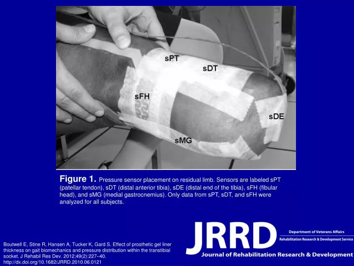
Figure 1. Pressure sensor placement on residual limb. Sensors are labeled sPT (patellar tendon), sDT (distal anterior tibia), sDE (distal end of the tibia), sFH (fibular head), and sMG (medial gastrocnemius). Only data from sPT, sDT, and sFH were analyzed for all subjects.

E N D
Figure 1. Pressure sensor placement on residual limb. Sensors are labeled sPT (patellar tendon), sDT (distal anterior tibia), sDE (distal end of the tibia), sFH (fibular head), and sMG (medial gastrocnemius). Only data from sPT, sDT, and sFH were analyzed for all subjects. Boutwell E, Stine R, Hansen A, Tucker K, Gard S. Effect of prosthetic gel liner thickness on gait biomechanics and pressure distribution within the transtibial socket. J Rehabil Res Dev. 2012;49(2):227–40.http://dx.doi.org/10.1682/JRRD.2010.06.0121