Download
1 / 107
1.07k likes | 1.09k Views
CARDIOVASCULAR DISEASE. Dariush Abtahi MD Anesthesiologist. Cardiovascular disease is the leading cause of death. Coronary artery disease, peripheral vascular disease, and risk for coronary artery disease increase operative risk.

E N D
CARDIOVASCULARDISEASE Dariush Abtahi MD Anesthesiologist
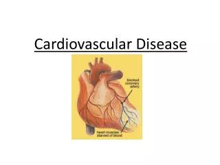
Cardiovascular disease is the leading cause of death. • Coronary artery disease, peripheral vascular disease, and risk for coronary artery disease increase operative risk.
Recent myocardial infarction, the presence of congestive heart failure, and aortic stenosis are the most common major risk factors.
In 40% of the adult patients who undergo surgery annually in the United States. • Increased rates of morbidity and mortality when coronary artery disease is present.
Routine preoperative cardiac evaluation: • physical examination • evaluation of exercise tolerance • cardiac symptoms • ECG
The most common symptoms: • shortness of breath with exercise in men and fatigue in women • angina • angina at rest • orthopnea • paroxysmal nocturnal dyspnea • dizziness or fainting
More specialized procedures: • ambulatory ECG monitoring (Holter monitoring) • exercise stress testing • transthoracic or transesophageal echocardiography • radionuclide ventriculography (determination of ejection fraction) • dipyridamole-thallium scintigraphy • cardiac catheterization • angiography
More specialized procedures: • NO MORE INFORMATION!
Patient History • Patients can remain asymptomatic despite 50% to 70% stenosis of a major coronary artery.
CARDIAC RESERVE • Limited exercise tolerance in the absence of significant pulmonary disease is the most striking evidence of decreased cardiac reserve.
Evidence of significant cardiac disease- inability to: • lie flat • awakening from sleep with angina or shortness of breath • angina at rest or with minimal exertion
If a patient can climb two to three flights of stairs without symptoms, cardiac reserve is probably adequate.
ANGINA PECTORIS • Stable: no change for at least 60 days.
Unstable: chest pain or shortness of breath produced with less than normal activity or at rest, or lasting for increasingly longer periods • Signal an impending myocardial infarction.
Silent myocardial ischemia (asymptomatic). • 70% of ischemic episodes- 15% of acute myocardial infarctions!
Women and diabetics have a more frequent incidence of painless myocardial ischemia and infarctions.
MYOCARDIAL INFARCTION • The incidence of perioperative myocardial reinfarction does not stabilize at 5% to 6% until 6 months after the prior myocardial infarction.
Elective surgery, especially thoracic, upper abdominal, or other major procedures are delayed for a period of 2 to 6 months after a myocardial infarction.
After 6 months, the 5% to 6% incidence of myocardial reinfarction is about 50 times more frequent than the 0.13%.
Most perioperative myocardial reinfarctions occur in the first 48 to 72 hours postoperatively.
Several factors influence the incidence of myocardial infarction in the perioperative period: • Intra-thoracic or intra-abdominal operations lasting longer than 3 hours.
Decrease the risk: • Intensive hemodynamic monitoring using an intra-arterial catheter • Prompt pharmacologic intervention or fluid infusion to treat physiologic hemodynamic alterations
CURRENT MEDICATIONS • Do Not Stop: beta-blockers, calcium channel blockers, nitrates, statins, or angiotensin-converting enzyme inhibitors in the perioperative period.
Electrocardiogram • ECG examined for: • (1) myocardial ischemia • (2) prior MI • (3) cardiac hypertrophy • (4) abnormal cardiac rhythm • (5) electrolyte abnormalities
The exercise ECG simulates events such as direct laryngoscopy, tracheal intubation, and surgical incision.
The resting ECG in the absence of angina pectoris may be normal despite extensive coronary artery disease.
Management of Anesthesia • Anesthesia care for patients with known coronary artery disease, or two risk factors for coronary artery disease should begin as soon as the patient is identified as needing surgery.
Risk Factors: age older than or equal to 60 years hypertension diabetes significant smoking history hyperlipidemia
Avoid: • persistent tachycardia • systolic hypertension • arterial hypoxemia • diastolic hypotension
Maintaining heart rate and systemic blood pressure within 20% of the awake values is commonly recommended.
Monitoring with an intra-arterial catheter facilitates the ability to maintain stable systemic blood pressures.
A single 1-minute episode of myocardial ischemia detected by 1-mm ST-segment elevation or depression increases the risk of cardiac events tenfold and the risk for death twofold.
Tachycardia for 5 minutes above 120 beats/min in the postoperative period can increase the risk of death tenfold.
MONITORING • Anticipation of problems and avoidance of potential disasters is a key component in successful anesthetic management in patients with cardiovascular disease. • Prophylactic therapy and more extensive monitoring reduce risk.
Rapid changes in hemodynamics can quickly lead to cardiac arrest • Monitoring can quickly identify those changes
When operations are completed, monitoring should be continued into the recovery room or intensive care unit (ICU).
Loss of a pulse oximeter signal: • hypoxia • inadequate arterial blood pressure
INDUCTION OF ANESTHESIA • Preoperative anxiety can lead to preoperative myocardial ischemia. • Patients should receive their routine medications except for oral hypoglycemic drugs.
Etomidate is a popular anesthetic to induce anesthesia (limited hemodynamic effects). • Propofol is popular, but the dose should be reduced to avoid hypotension with induction.
Myocardial ischemia may accompany the tachycardia and hypertension that results from the stimulation of direct laryngoscopy.
Adequate anesthesia and a brief duration of direct laryngoscopy is important in minimizing the magnitude of these circulatory changes.
MAINTENANCE OF ANESTHESIA • Avoiding tachycardia with the use of long-acting beta-blockers is more important than anesthetic choice.
A regional anesthetic is an excellent technique in patients with coronary artery disease.
Decreases in systemic blood pressure associated with a regional anesthetic that are more than 20% of the pre-block value probably should be treated with an intravenous infusion of crystalloid solutions or a vasoconstrictor such as phenylephrine.
NEUROMUSCULAR BLOCKING DRUGS • Cisatracurium do not evoke histamine release and associated decreases in systemic blood pressure, even with the rapid intravenous administration of large doses.
The systemic blood pressure lowering effects of atracurium are usually modest, especially if the drug is injected over 30 to 45 seconds to minimize the likelihood of drug-induced histamine release.
Pancuronium increases heart rate and systemic blood pressure, but these changes are usually less than 15% above pre-drug values, making this drug a possible choice for administration to patients with coronary artery disease.