Download
1 / 25
310 likes | 484 Views
Hemolytic Disease of the Fetus/Newborn and Intrauterine RBC Transfusion. Raina Raquel Flores, M.D. 3/7/14. Outline. Brief review: Hemolytic disease of the fetus and newborn (HDFN) Intrauterine RBC transfusion Indications Blood product selection Transfusion technique Volume to transfuse

E N D
Hemolytic Disease of the Fetus/Newborn and Intrauterine RBC Transfusion Raina Raquel Flores, M.D. 3/7/14
Outline • Brief review: Hemolytic disease of the fetus and newborn (HDFN) • Intrauterine RBC transfusion • Indications • Blood product selection • Transfusion technique • Volume to transfuse • Complications • Fetal IVIG Therapy
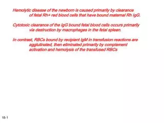
HDFN • Destruction of fetal/newborn RBCs by antibodies (IgG) produced by the mother • Maternal antibodies to ABO antigens are most common cause of HDFN (mild) • Majority of severe HDFN due to anti-Rh(D) antibodies • Anti-c, E, and C antibodies also clinically significant • Of the non-Rh system antibodies, anti-Kell is most common cause of HDFN
Pathogenesis Active IgG transport (2nd trimester)
Pathogenesis Fetal anemia Marrow stimulated to release immature RBCs – erythroblastosis fetalis Fetal RBCs coated in maternal IgG Extravascular hemolysis in spleen High output cardiac failure with ascites, effusions, edema – hydrops fetalis Decreased hepatic plasma protein production Extramedullary hematopoiesis in liver
Sir William Liley (1929 - 1983) In 1961, demonstrated that amniotic fluid bilirubin corresponds to degree of fetal anemia → Liley curve Observed improved anemia in sickle cell patients after peritoneal infusion with RBCs In 1963, Liley reports first successful intrauterine transfusion for HDFN “Intrauterine Transfusion of Foetus in Haemolytic Disease” Br Med J 1963;5365:1107e9.
Intrauterine RBC Transfusion • Gold standard for treatment of severe fetal anemia • RBC alloimmunization • Anti-D, anti-Kell, and anti-c • Parvovirus infection • Chronic fetomaternal hemorrhage • Inherited RBC disorders • Congenital dyserythropoietic anemia, alpha thalassemia • Complications after twin-twin transfusion syndrome
Assessing Fetal Anemia Assessing severity of fetal anemia: Middle cerebral artery-peak systolic velocity (MCA-PSV) Anemic fetus preserves O2 delivery to the brain by increasing cerebral flow Fetal blood sampling 1-2% risk of fetal loss Reserved for cases with increased MCA-PSV Amniotic fluid spectral analysis (Liley curve) N Engl J Med. 2000;342(1):9.
When to Initiate Intrauterine Transfusion (IUT) Indications: Cordocentesis blood sample with: Hgb < 10 g/dL Hgb level > 7 below the mean for GA Hgb < 2 SD below the mean for GA Hct <30% or < 2 SD below the mean for GA Fetal hydrops noted on ultrasound Amniotic fluid ΔOD 450 nm results in high zone II or zone III N Engl J Med. 2000;342(1):9
RBC Product Selection Requirements: Type O Rh(D)-, antigen negative, crossmatch compatible CMV seronegative Leukoreduced Irradiated Hemoglobin S negative Fresh (less than 7 days old) Washed and tightly packed Hct 75-85% Considerations: Donor vs. maternal RBCs? Seminars in Fetal & Neonatal Medicine (2007) 12, 432e438
Donor vs. Maternal RBCs • 25% of mothers undergoing IUT produce additional RBC antibodies due to exposure to antigens on donor blood • Use of autologous blood from mother decreases risk of sensitization • Donation may be limited by Hgb status of the mother • Possible decrease in frequency of IUT in neonates who receive maternal blood Am J Obstet Gynecol. 1997;177(4):753.
Intraperitoneal Transfusion • Indirect access to fetal circulation • RBCs slowly absorbed through diaphragmatic lymphatic system (over 7 to 10 days) • Least effective in the hydropic fetus • May be preferred if fetal vasculature inaccessible Fetal Diagn Ther. 2008;23(2):159.
Direct Vascular Access • Correction of fetal anemia is rapid • More effective than IPT in the hydropic fetus • Intravascular transfusion (IVT) improves survival two-fold in the hydropic fetus • Overall, higher rate of survival with IVT compared to IPT Am J Obstet Gynecol. 1991;165(4 Pt 1):1020.
Umbilical Cord Transfusion • Preferred method in North America • Target: umbilical vein at site of cord insertion into placenta • Stable • Less fetal bradycardia Am J Anat. 1971;132(1):53.
Intrahepatic Umbilical Vein Transfusion • Preferred method in Europe • Target: intrahepatic portion of the umbilical vein • Low incidence of fetal bradycardia • Fetal pain → fetal movement → organ trauma Lancet. 1994;344(8915):77. Am J Obstet Gynecol. 2005;192(1):171
Combined Approach • IPT followed by IVT • If transfusion is required prior to 18-20wks gestation • IVT followed by IPT • More stable fetal hematocrit between procedures • Slow absorption of RBCs from intraperitoneal reservoir • Longer interval between procedures → fewer total IUTs Fetal Ther. 1989;4(2-3):126
IUT Volume Determination • Target Hct depends on gestational age • Less than 24 weeks gestation: • 1st IUT → Hct ≤ 25% • 2nd IUT (48 hours later) → Hct ~ 40% • 3rd IUT (7 to 10 days later) → Hct ~ 40% • Greater than 24 weeks gestation: Hct 40 to 50% • Intraperitoneal transfusion (IPT) volume: Volume to transfuse (ml) = (GA in weeks – 20) x 10 ml • Intravascular transfusion volume: Fetoplacental volume (FPV) (ml) = fetal weight (g) x 0.14 Volume to transfuse (ml) = [FPV x (Goal Hct – Initial Hct)] / Donor unit Hct Obstet Gynecol. 1992;79(3):390
Timing of Transfusion in Alloimmunization • After 1st IUT, Hct will drop by 1% per day if not hydropic and 1.88% per day if hydropic • Fetal erythropoiesis suppressed after 2 to 3 transfusions → lengthened time interval between subsequent procedures • Can empirically transfuse. . . • 10 days after 1st transfuion • 2 weeks after 2nd transfusion • 3 weeks after 3rd transfusion • Transfusions should continue up to 34-35 weeks gestation
Complications of IUT • Most common complication of IPT is infusion in the wrong location • For intravascular transfusions • Procedure-related (PR) fetal loss = 1 to 2% per procedure • Overall risk of PR complications = 3% per procedure • Emergency c/s • Infection • Bleeding from puncture site • Premature rupture of membranes • Arterial puncture • Bradycardia or tachycardia American Journal of Obstetrics and Gynecology (2005) 192, 171e7
Outcomes of IUT • Survival: • 89% survival after one IUT • Survival of hydropic fetus is lower than non hydropic fetus • Lower survival if IUT needed at less than 20 weeks • Neurologic: • Normal neurologic outcome expected in 90% of surviving infants, regardless of hydropic status at time of 1st IUT • Hydropic fetus more likely to severe developmental delay or cerebral palsy Obstet Gynecol. 1996;88(1):137 Am J Obstet Gynecol. 2012;206(2):141.e1
IVIG Therapy • In Rh(D) and ABO HDFN babies, postnatal IVIG reduces need for exchange transfusion for hyperbilirubinemia • IVIG may block antibody receptors on RBCs • Maternal IVIG administration during pregnancy for management of alloimmune HDFN • Expensive • Questionable efficacy Arch Dis Child Fetal Neonatal Ed. 2003;88(1):F6
Direct Fetal Immunoglobulin Administration?? • “Fetal Intravenous Immunoglobulin Therapy in Rhesus Hemolytic Disease” • 4 cases of severe Rh(D) HDFN treated with fetal IVIG • Decreased frequency of IUT and eliminated need for postnatal transfusion • “Fetal intraperitoneal injection of immunoglobulin diminishes alloimmune hemolysis” • Case of anti-M HDFN successfully managed with 4 IG treatments (2 g per-kg fetal body weight) • Healthy baby delivered at 38 weeks without IUT, exchange transfusion, or phototherapy J Perinatol. Apr 2011; 31(4): 289–292. Gynecol Obstet Invest 2007;63:176–180