Download
1 / 26

E N D
Ascaris lumbricoides is one of the largest and most common parasites found in humans. The adult females of this species can measure up to 45 cm long (males are generally shorter), and it is estimated that 25% of the world's population is infected with this nematode. The adult worms live in the small intestine and eggs are passed in the feces. A single female can produce up to 200,000 eggs each day!
About two weeks after passage in the feces the eggs contain an infective larval or juvenile stage, and humans are infected when they ingest such infective eggs. The eggs hatch in the small intestine, the juvenile penetrates the small intestine and enters the circulatory system, and eventually the juvenile worm enters the lungs. In the lungs the juvenile worm leaves the circulatory system and enters the air passages of the lungs. The juvenile worm then migrates up the air passages into the pharynx where it is swallowed, and once in the small intestine the juvenile grows into an adult worm. Why does Ascaris undergo such a migration through the body to only end up where it started? Such a migration is not unique to Ascaris, as its close relatives undergo a similar migration in the bodies of their hosts
large mass of Ascaris lumbricoides that was passed from the intestinal tract. The ruler at the bottom of the image is 4 cm
Ascaris infections in humans can cause significant pathology. The migration of the larvae through the lungs causes the blood vessels of the lungs to hemorrhage, and there is an inflammatory response accompanied by edema. The resulting accumulation of fluids in the lungs results in "ascaris pneumonia," and this can be fatal. The large size of the adult worms also presents problems, especially if the worms physically block the gastrointestinal tract. Ascaris is notorious for its reputation to migrate within the small intestine, and when a large worm begins to migrate there is not much that can stop it. Instances have been reported in which Ascaris have migrated into and blocked the bile or pancreatic duct or in which the worms have penetrated the small intestine resulting in acute (and fatal) peritonitis. Ascaris seems to be especially sensitive to anesthetics, and numerous cases have been documented where patients in surgical recovery rooms have had worms migrate from the small intestine, through the stomach, and out the patient's nose or mouth.
In the lungs, the larvae break out of the pulmonary capillaries into the air sacs, ascend into the throat and descend to the small intestine again where they grow. Molting (ecdysis) occurs at various points along this path and, typically for roundworms, the male and female adults in the intestine are 5th-stage nematodes. Vague digestive tract discomfort sometimes accompanies the intestinal infection, but in small children with more than a few worms there may be intestinal blockage because of the worms' large size. Not all larval or adult worms stay on the path that is optimal for their development; those that wander may locate in diverse sites throughout the body and cause complications. Chemotherapy with anthelmintics is particularly likely to cause the adult worms in the intestinal lumen to wander; a not unusual escape route for them is into the bile duct which they may occlude. The larvae of ascarid species that mature in hosts other than humans may hatch in the human intestine and are especially prone to wander; they may penetrate into tissues and locate in various organ systems of the human body, perhaps eliciting a fever and diverse complications.
What are the signs and symptoms of an ascaris infection? Most people have no symptoms. If you are heavily infected, you may have abdominal pain. Sometimes, while the immature worms migrate through the lungs, you may cough and have difficulty breathing. If you have a very heavy worm infection, your intestines may become blocked. Ascarid eggs are found in the soil. Infection occurs when a person accidently ingests (swallows) infective ascarid eggs. Once in the stomach, larvae (immature worms) hatch from the eggs. The larvae are carried through the lungs then to the throat where they are then swallowed. Once swallowed, they reach the intestines and develop into adult worms. Adult female worms lay eggs that are then passed in feces; this cycle will take between 2-3 months.
The infective stage is the egg containing an L2 larva, which is ingested by the host and hatches in the duodenum. In lungs- two moults Move up respiratory tree to pharynx- swallowed Most J3s cannot stand gastric juices, J4s can- pass to small intestine- produce eggs in ~2 months Environmental contamination via indiscriminate defecation Eggs resistant-2% formalin, 50% solutions of acid (This resistance is due to a lipid layer in the egg shell which contains lipids unique to the ascaris called ascarocides. They also have an extra uterine layer or coat, which makes the eggs very sticky and aids transmission). Eggs may remain in soil for 5-10 years Children infected – why? Study: found eggs on uncooked vegetables, nasal mucous (wind-borne), on banknotes Worms commonly over dispersed- few people harboring lots- these people tend to predisposed to infection- after clearance often reinfected- behavioural or biological?
Juveniles cause hemorrhages as they break out of lungs- can cause pools of blood, edema, clogging of lung spaces, accumulations of white blood cells, - Ascaris pneumonitis. Large areas of lungs may be compromised and secondary infections set in. Nematodes feed little on blood or mucosa- feed mainly on liquid contents of intestine. Can cause malnutrition, underdevelopment. Abdominal pains, rashes, asthma, and restlessness thought to be due to allergic responses to worm metabolites Massive infections- block intestine. Many times worms cause no problems, other times they gather in knots and block lumen. Some drugs can induce this. Wandering adults: some simply penetrate intestine, peritonitis often fatal. May wander down to appendix, exit anus, upwards to stomach- irritate host- vomit adult.
Secondary complications can arise with ascaris infections because sometimes, when the worms are undergoing this migration they appear to get lost and start wandering through other organs such as the brain, bile duct, pancreas or appendix. Even the adult worms can start to wander, often as a result of overcrowding as a result of a heavy infection, or an absence of male worms and even in response to certain anthelmintic treatments. When this occurs, worms have been known to exit the host via the anus, mouth and nose.
Although ascaris has only a single host and it is found in the small intestine, its life cycle is far from simple. It has been suggested that from an evolutionary perspective that ascaris originally had two hosts and has secondarily lost its intermediate host. It is this rather bizarre migration which the parasite undergoes, to effectively get back to where it started, which has led to the suggestion that perhaps originally that this migratory path originally took place in an intermediate host.
Ascarid evolution Why would Ascaris enter via the mouth and then migrate through the body- lungs-coughed up- just to get back to the intestine. This is not “logical” Often considered to be a multi-host system compressed into one host- with the same host acting as intermediate and definitive hosts. Consider: Primitive ascarids were free living marine worms Terrestrial worms arose secondarily Indirect life cycle is widespread in marine and terrestrial species What we see may be a snapshot in an evolutionary continuum
Toxocara canis In most cases, Toxocara infections are not serious, and many people, especially adults infected by a small number of larvae may not notice any symptoms. The most severe cases are rare, but are more likely to occur in young children, who often play in dirt, or eat dirt contaminated by dog or cat stool. The most common Toxocara parasite of concern to humans is T. canis, which puppies usually contract from the mother before birth or from her milk. The larvae mature rapidly in the puppy’s intestines; when the pup is 3 or 4 weeks old, they begin to produce large numbers of eggs that contaminate the environment through the animal’s stool. The eggs soon develop into infective larvae.
There are two major forms of toxocariasis: 1) Ocular larva migrans (OLM): Toxocara infections can cause OLM, an eye disease that can cause blindness. OLM occurs when a microscopic worm enters the eye; it may cause inflammation, cause lesions, and formation of a scar on the retina. Each year more than 700 people infected with Toxocara experience permanent or partial loss of vision. 2) Visceral larva migrans (VLM): Heavier, or repeated Toxocara infections, while rare, can cause VLM, a disease that causes swelling of the body’s organs or central nervous system. The migrating larvae invade multiple tissues (liver, heart, lungs, brain, muscle) and cause various symptoms including fever, anorexia, weight loss, cough, wheezing, rashes, hepatosplenomegaly, and hypereosinophilia. In this parasitic disease, diagnosis does not rest on identification of the parasite. Since the larvae do not develop into adults in humans, a stool examination would not detect any Toxocara eggs. However, the presence of Ascaris and Trichuris eggs in feces, indicating fecal exposure, increases the probability of Toxocara in the tissues. A presumptive diagnosis rests on clinical signs, history of exposure to puppies, laboratory findings (including eosinophilia), and the detection of antibodies to Toxocara.
Biology of Toxocara canis Toxocara canis is a remarkable nematode parasite, commonly found in dogs but able to invade a wide range of other hosts including humans. It displays an array of striking features which excite interest: a tissue-dwelling phase which can endure many years; an ability to cross the placenta and infect unborn pups; a tropism for neurological tissue survival in vitro for many months in serum-free medium; the secretion of a set of biologically active glycoproteins in vivo and in vitro; the possession of a surface glycocalyx which is jettisoned under immune attack. For all these reasons, T.canis presents an important model system for parasitic nematodes. In addition, toxocariasis can cause significant pathology in humans as well as dogs, and elimination of this infection would be a highly desirable goal.
Life cycle • Eggs hatch in seawater • Larvae are eaten by crustaceans • The crustaceans are eaten by a fish • The human becomes an incidental host. Anisakis infects human, by eating raw or undercocked fish or crustaceans.
http://www.clinicasubiza.com/data/enfermedades/anisalis2.htm research.kahaku.go.jp/.../kaisei/ hp-9/ikikata/sukeso.html ucdnema.ucdavis.edu/imagemap/
Anisakis (simplex) A roundworm living in the stomach The roundworm has a layer which protects it from the gastric acids, which makes it possible for the worm to burrow into the gastric wall. Causes Anisakisis: infection of the gastrointestinal tract caused by the consumption of raw or undercooked seafood containing larvae of the nematode Anisakis (simplex) Can be found in areas where fish is consumed raw, lightly picked or salted. Example Japan, Scandinavia, Netherlands and along the pacific coast of South America (but no longer by students of BISC 318!)
Symptoms Just after a few hours of eating the infected larvae, the patient may have abdominal pain and vomiting and nausea may occur. • Abdominal pain: • - Sudden severe pain • Nausea • Vomiting • Diarrhea • Loss of appetite • Acute allergic symptoms: • Rash • Throat swelling • Low blood pressure • (the allergic symptoms; with or without gastrointestinal symptoms)
Diagnosis Gastroscopic examination (the larva are visualized and removed) Histopathologic examination (tissue removed at biopsy or during surgery) Diagnosis is usually made by upper endoscopy, Exploratory laparotomy (small intestine, cecum, colon)
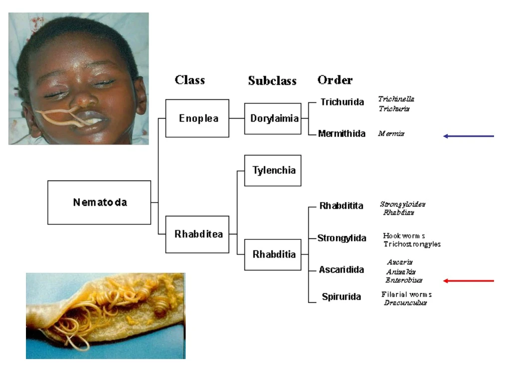
Nematodes: • Mermithids: always kill their invertebrate hosts, free living and parasitic stage, transmission via ingested eggs or penetration by juveniles • 2) Ascarids have no free living stage. Eggs are released into environment. Immature stages go on migratory walks in the host before returning to the GI tract.