Download
1 / 29
290 likes | 593 Views
Joints. Articulations of bones Functions of joints Hold bones together Allow for mobility Ways joints are classified Functionally Structurally. Functional Classification of Joints. Synarthroses Immovable joints Amphiarthroses Slightly moveable joints Diarthroses

E N D
Joints • Articulations of bones • Functions of joints • Hold bones together • Allow for mobility • Ways joints are classified • Functionally • Structurally
Functional Classification of Joints • Synarthroses • Immovable joints • Amphiarthroses • Slightly moveable joints • Diarthroses • Freely moveable joints
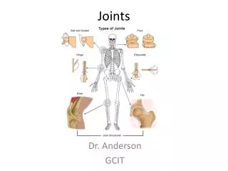
Structural Classification of Joints • Fibrous joints • Generally immovable • Cartilaginous joints • Immovable or slightly moveable • Synovial joints • Freely moveable
Summary of Joint Classes [Insert Table 5.3 here] Table 5.3
Fibrous Joints • Bones united by fibrous tissue • Example: • Sutures • Syndesmoses • Allows more movement than sutures • Example: Distal end of tibia and fibula
Fibrous Joints Figure 5.28a–b
Cartilaginous Joints • Bones connected by cartilage • Example: • Pubic symphysis • Intervertebral joints
Cartilaginous Joints Figure 5.28c–e
Synovial Joints • Articulating bones are separated by a joint cavity • Synovial fluid is found in the joint cavity
Synovial Joints Figure 5.28f–h
Features of Synovial Joints • Articular cartilage (hyaline cartilage) covers the ends of bones • A fibrous articular capsule encloses joint surfaces • A joint cavity is filled with synovial fluid • Ligaments reinforce the joint
Structures Associated with the Synovial Joint • Bursae—flattened fibrous sacs • Lined with synovial membranes • Filled with synovial fluid • Not actually part of the joint • Tendon sheath • Elongated bursa that wraps around a tendon
The Synovial Joint Figure 5.29
Types of Synovial Joints Figure 5.30a–c
Types of Synovial Joints Figure 5.30d–f
Inflammatory Conditions Associated with Joints • Bursitis—inflammation of a bursa usually caused by a blow or friction • Tendonitis—inflammation of tendon sheaths • Arthritis—inflammatory or degenerative diseases of joints • Over 100 different types • The most widespread crippling disease in the United States
Clinical Forms of Arthritis • Osteoarthritis • Most common chronic arthritis • Probably related to normal aging processes • Rheumatoid arthritis • An autoimmune disease—the immune system attacks the joints • Symptoms begin with bilateral inflammation of certain joints • Often leads to deformities
Clinical Forms of Arthritis • Gouty arthritis • Inflammation of joints is caused by a deposition of uric acid crystals from the blood • Can usually be controlled with diet
Developmental Aspects of the Skeletal System • At birth, the skull bones are incomplete • Bones are joined by fibrous membranes called fontanels • Fontanels are completely replaced with bone within two years after birth
Ossification Centers in a 12-week-old Fetus Figure 5.32
Skeletal Changes Throughout Life • Fetus • Long bones are formed of hyaline cartilage • Flat bones begin as fibrous membranes • Flat and long bone models are converted to bone • Birth • Fontanels remain until around age 2
Skeletal Changes Throughout Life • Adolescence • Epiphyseal plates become ossified and long bone growth ends • Size of cranium in relationship to body • 2 years old—skull is larger in proportion to the body compared to that of an adult • 8 or 9 years old—skull is near adult size and proportion • Between ages 6 and 11, the face grows out from the skull
Skeletal Changes Throughout Life Figure 5.33a
Skeletal Changes Throughout Life Figure 5.33b
Skeletal Changes Throughout Life • Curvatures of the spine • Primary curvatures are present at birth and are convex posteriorly • Secondary curvatures are associated with a child’s later development and are convex anteriorly • Abnormal spinal curvatures (scoliosis and lordosis) are often congenital
Skeletal Changes Throughout Life Figure 5.16
Skeletal Changes Throughout Life • Osteoporosis • Bone-thinning disease afflicting • 50% of women over age 65 • 20% of men over age 70 • Disease makes bones fragile and bones can easily fracture • Vertebral collapse results in kyphosis (also known as dowager’s hump) • Estrogen aids in health and normal density of a female skeleton
Skeletal Changes Throughout Life Figure 5.34
Skeletal Changes Throughout Life Figure 5.35