Download
1 / 24
240 likes | 314 Views
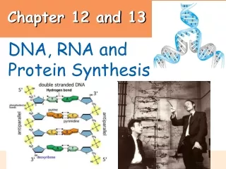
Chapter 12 and 13. 0. DNA, RNA and Protein Synthesis. Discovery of the Role of DNA. A. 1928 - Frederick Griffith discovers transformation in bacteria : * discovered that “something” was able to transform harmless (non – virulent) bacteria into harmful (virulent).

E N D
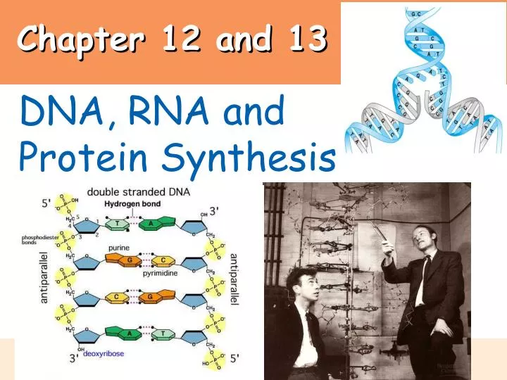
Chapter 12 and 13 0 DNA, RNA and Protein Synthesis
Discovery of the Role of DNA A. 1928 - Frederick Griffith discovers transformation in bacteria : * discovered that “something” was able to transform harmless (non – virulent) bacteria into harmful (virulent)
Discovery of the Role of DNA (cont’d) • 1944 -Oswald Avery and colleagues show that DNA can transform bacteria • 1952 - Alfred Hershey and Martha Chase use bacteriophage to confirm that DNA is the genetic material
Hershey-Chase Experiment: Infected cells make more virus by injecting their DNA animation 1
Discovery of the Role of DNA (cont’d) D. 1953 - James Watson and Francis Crick propose a structural model for the DNA molecule Based On: X-Ray crystallography images prepared by Maurice Wilkins and Rosalind Franklin Chargraff’s Rule: # of Adenines = # of Thymines # Guanines = # of Cytosines
DNA and RNA are Polymers of Nucleotides • Both are nucleic acids made of long chains of nucleotide monomers • A nucleotide (building block of a nucleic acid) has 3 parts: • A phosphate (PO4-) group that is negatively charged • A 5-Carbon sugar (deoxyribose in DNA or ribose in RNA) • A nitrogen-containing base
Thymine (T) Cytosine (C) Adenine (A) Guanine (G) DNA (deoxyribonucleic acid) bases: purines pyrimidines • Pyrimidines: single ring bases • Purines: double ring bases • Complimentary binding pattern: • Adenine + Thymine (share 2 hydrogen bonds) • Cytosine + Guanine (share 3 hydrogen bonds)
RNA: ribonucleic acid Similar to DNA except: • Sugar in RNA = ribose • Base “uracil” instead of thymine • Single stranded Figure 10.2C, D
Twist The Structure of DNA • Two polynucleotide strands wrapped around each other in a double helix • A sugar-phosphate backbone • Steps made of hydrogen-bound bases (A=T, C = G)
DNA REPLICATION: Starts with the separation of DNA strands • Enzymes use each strand as a template to assemble new nucleotides into complementary strands…“semi-conservative” (Meselson & Stahl 1958) • Portions to be replicated must untwist first
DNA segments unwind 2. Helicase splits H bonds between bases, unzip DNA 3. Binding proteins keep unzipped DNA apart (Single Stranded Binding Proteins) 4. Primase makes a short RNA primer because DNA polymerase can only extend a nucleotide chain, not start one. 5. DNA polymerase adds new nucleotides to the 3’ end of daughter strand that are complimentary to the parent strand 6. RNase H cuts out original primers 7. DNA polymerase fills in gap of removed primers 8. DNA ligase glues S/P backbone where needed DNA replication begins at specific sites on double helix replication forks Animation/tutorial 9. Two identical double helices • Topoisomerase: prevents further coiling at replication fork
A Structural Problem with DNA Replication • Each strand of the double helix is oriented in the opposite direction (“anti-parallel”) • “prime” #’s refer to carbons in the sugar • At one end, the 3’ carbon has an (OH) and at the opposite, a 5’ carbon has the PO4- • Why does this matter? DNA polymerase can only add nucleotides to the 3’ end. A daughter strand can only grow from 5’ 3’ • Therefore, only one daughter strand is made continuously (leading strand) • The other strand (lagging strand) is made in a series of short pieces (Okazaki fragments), later connected by DNA ligase animation Animation/tutorial
When DNA can repair mistakes and when it can’t DNA Repair enzymes work like a spell checker • Cut out wrong sequences • Undamaged strand is template • Only 2 or 3 stable changes per year : some severe, others are not • Mutations Inheritable changes occur in gametogenesis • Now the “wrong” sequences are copied • Ex: cystic fibrosis (CF): a deletion of 3 nucleotides in a certain gene • Ex: sickle cell anemia: one nucleotide substitution in the hemoglobin gene • Mutagen: a mutation causing substance (can break DNA) • Ex: X-Rays, radioactivity, nicotine
Protein Synthesis: the transfer of information from: DNA RNA Proteins “gene expression”: A gene is a linear sequence of many nucleotides. 3 Types: • Structural genes: have info to make proteins • Regulatory genes: are on/off switches for genes • Genes that code for tRNA, rRNA, histones DNA vs. RNA • single stranded • A U C G • ribose sugar • 3 types of RNA: • messenger, transfer, ribosomal • double stranded • A T C G • deoxyribose sugar mRNA (messenger): copies DNA’s message in nucleus brings it to cytoplasm tRNA (transfer): carries amino acids to mRNA so protein can be made rRNA (ribosomal): major part of the ribosome. Helps link amino acids from tRNA’s together assemble protein
Transcription: The DNA of the gene is transcribed into mRNA Translation: decoding the mRNA and assembling the protein Protein Synthesis is Two Steps:
Transcription: Eukaryote DNA sequence (message for protein) is transcribed by mRNA Only one strand (non-coding strand) is needed as a template Steps: RNA polymerase splits H bonds in DNA section RNA polymerase travels along non-coding strandof DNA. RNA nucleotides join in a complimentary pattern (A=U, C=G) A termination signal is reached, transcription is over mRNA strip detaches from DNA, DNA helix closes up mRNA is processed: Introns are cut out, Exons are glued together, cap and tail are added. Mature mRNA leaves nucleus through pores cytoplasm for next step
Translation: the synthesis of proteins using mRNA, tRNA and ribosomes The Genetic Code: the language in which instructions for proteins are written in the base sequences Each triplet of mRNA bases is a “codon” because it will “code” for 1 amino acid Ex: AUG GUC CCU AAU CCU Met – Val – Pro – Asn – Pro Original coding strand of DNA (the actual gene): ATG GTC CCT AAT CCT Only difference: U is substituted for T Use the Genetic Code chart to “decode” mRNA message
Nearly all organisms use exactly the same genetic code More than one codon for most amino acids = degenerate nature…a change (mutation) in gene does not always mean a different amino acid. what does CAU code for? ACU? UAU? GCC? how many codons for Leu? what is special about AUG and it’s amino acid, Methionine? what is special about UAA, UAG, and UGA? The Genetic Code is the Rosetta Stone of Life
An exercise in translating the genetic code: Step 1: fill in corresponding DNA bases to dark blue strand (non-coding) Step 2: Transcribe the dark blue strand into mRNA (pink) Step 3: Translate the codons into correct amino acids (use chart) G A A A T G T G T T T A Coding strand (gene) transcription translation
An exercise in translating the genetic code: answers Step 1: fill in corresponding DNA bases to dark blue strand (non-coding) Step 2: Transcribe the dark blue strand into mRNA (pink) Step 3: Translate the codons into correct amino acids (use chart)
Need: tRNAs and ribosomes (rRNA) tRNA: single stranded RNA, folded up 2 parts: anticodon and aa attachment site How Does Translation Happen? • Ribosome: 2 protein subunits and ribosomal RNA • allows aa’s to attach by making peptide bonds • travels along mRNA strip, tRNA’s join and bring correct amino acids • 3 sites on ribosome: • A site – where new tRNA’s and amino acids join • P site – where protein is growing • E site – where empty tRNA’s exit ribosome • Translocation: as ribosome moves, tRNA’s move from A site to P site. “A” site is now open for new tRNA with attached amino acid to join animation
Mutations can change the message of genes • Mutations: • changes in DNA base sequence • caused by errors in DNA replication, recombination, or by mutagens • substituting, inserting, or deleting nucleotides also alters a gene “point mutation”…may or may not alter amino acid sequence “frame-shift mutation”…most devastating to protein structure
MUTANTS – • Mutant Animals!