Download
1 / 52
520 likes | 662 Views
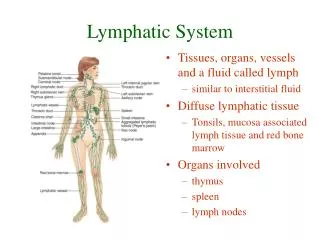
Lymphatic System. 1-The lymphatic tissues which are: A-Lymph nodes B-Spleen C-Scattered lymphatic nodules D-Tonsils 2-The Lymphatic vessels: the lymphatic capillaries and lymph vessels;the carry the lymph. Formation of lymph.

E N D
Lymphatic System • 1-The lymphatic tissues which are: • A-Lymph nodes • B-Spleen • C-Scattered lymphatic nodules • D-Tonsils • 2-The Lymphatic vessels: the lymphatic capillaries and lymph vessels;the carry the lymph
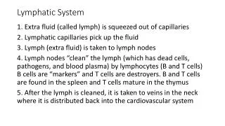
Formation of lymph • Tissue fluid which is filtered from the tissues and from the blood capillaries around the cells is drained by blind-ended lymphatic capillaries • The fluid when enters the lymphatic vessels called lymph
The lymph is filtred in lymph nodes&nodules • Lymph flows in one direction inside the lymphatic capillaries • The filtred lymph go again to the blood stream through alarge lymphatic vessels called thoracic duct
LYMPHATIC SYSTEM LYMPHATIC CAPILLARY artery vein lymphatic blood capillary venule capillaries LYMPHATIC SINUSOID arteriole lymphatic sinusoid
Differences between blood capillaries & lymphatic capillaries
Structures Of Lymph vessels • Large lymphatic vessels are formed of intima,media, adventitia • Similar to venis,they also have valves
Lymph nodes • Shape&function • Kidney shaped organs present along the course of lymphatic vessels • Function: filter the lymph form any organisme or forign body • Contain lymphocytes,macrophages,killer cells&plasma cells
Site of lymph nodes:present in groups in the axilla,neck,groin,thorax,abdomen7popliteal&cubital areas • Size; small,large in abnormal change afferent lymph vessels; bring lymph to lymph nodes • Efferent lymph carry lymph away from lymph nodes
Structures of lymph node • Is formed of C.T.stroma&parenchyma of: • Sroma • C.T. framework the lymph node include • 1-C.T.capsule • 2-C.T.trabeculae • 3-Reticular C.T.network
Capsule • Collagenous&elastic C.T.fibres separated by C.T. cells,smooth muscle are present at the hilum of lymph nodes • Trabeculae:formed of C.T. cells&fibers,divid the cortex of lymph node into compartment
3-Reticular Network: • Fine network of reticular C.T. formed of dendretic reticular cells&reticular fibres • Condensed more in cortex than in the medulla stained by silver stain
Cells of the stroma of the lymph node • A-Dendrtic Reticular Cells: they are branched cells with multiple cytoplasmic processes • Non-phagocytic cells,they are present in large numbers near B-lymphocytes
B-Macrophages cells: they are branched cells with large nuclei • They phagocytose foreign bodies,they are antigen presenting cells,secrete interleukin-I which regulates proliferation of lymphocytes • C-Fibroblast cells:they are present mainly around blood&lymph vessels
The parenchymal of lymph node • Parenchyma are functioning cells of the lymph node • Cortex;localized in the center of lymph nodes,irregular condensedation • The cortex of the lymph node contains • 1-cortical lymphatic nodules • 2-cortical lymphatic sinuses
1-The cortical lymphatic nodule • are of two types • a-primary lymphatic nodules • b-secondary lymphatic nodules
A-primary lymphatic nodules of follicles(aggregation of lymphocytes without germinal center) • -they are rounded,oval • -present under the capsule of lymph node • Aggregates of small lymphocytes • When the primary lymphatic nodules are exposed to infections or any antigens,small lymphocyte develop into activated
medium sized lymphocytes,newly formed activated lymphocytes aggregates in the center of the primary lymphatic nodules to form germinal centers&primary nodule changed into secondary lymphatic nodule
B-The secondary lymphatic nodules • Formed of the following cells • A-activated medium sized B-lymphocytes • B-T-lymphocytes¯ophages • C-Dendrtic reticular cells
2-The cortical lymphatic sinuses • Spaces which are present between the covering capsule&cortical lymphatic follicles • They are lined with flat endothelial cells • The cortical sinuses contain B-lymphocytes,macrophages&plasma cells
The Medulla of lymph node • Formed of • 1-Medullary lymphatic cord • -are irregular collections of lymphocyes&plasma cells • -containous with the cortical follicles • -seperated from each other by medullary lymphatic sinuses
2-Medullary lymphatic sinuses • -irregular wide spaces between the medullary cords • -lined with flat endothelial cells • -contain free lymphocytes,macrophages and some plasma cells
Circulation of lymph inside the lymph node • -lymph enters the lymph node by afferent lymph vessels through its cortex • -the lymph is filted through the cortical&medullary sinuses then the lymph leaves the node through efferent lymph vessels at is hilum
Blood supply of lymph node • Arteries enter the lymph nodes at hilum,branches pass to the cortex where they branch to form arterial capillaries • The venous capillaries:desend from the cortex to form post-capillary venules which lined by simple cubical cells collected veins leave the node at the hilum
Cells present in the lymph nodes • 1-B-Lymphocytes:common cells in both cortexand medulla • 2-T-lymphocytes: present in thymus dependent zones • 3-Macrophages:present inwhole stroma of lymph node • 4-Plasmablasts:present in cortex&medulla
6-Endothelial cells:lining the cortical&medullary lymph • 7-plasma cells;present in cortex and medulla of lymph node • 8-Endothelial cells:lining the cortical and medullary lymphatic sinuses
Function of lymph nodes • 1-filtration of lymph • 2-formation of lymphocytes • 3-production of antibodies formation of these immunoglobalin IgG,IgA,IgE,IgD
Spleen • -single intra-abdomenal haemolymphatic organ • -general filter for circulating blood • -has no afferent lymph vessels it has only efferent lymph vessels • Structures of spleen • Formed of C.T. stroma&parenchyma of lymphoid tissue
Stroma of spleen • C.T. framework which includes • A-capsule, • B-Trabeculae • C-Reticular C.T.
1-Capsule:covered peritoneum • Formed of collagenous&elastic C.T.fibres&fibroblast cells • The capsule is rich in smoth muscle • 2-trabeculae:formed of C.T.cells and fibres • The trabeculae is tich in smooth muscle • 3-Reticular network:made up of reticular fibres and cells • It can stained brown with silver stain • The reticular network is more condensed in the white pulp than red pulp
The Parenchyma • Composed of white&red pulp • White pulp or (malpighian corpuscles) • They are rounded or elongated lymphatic nodules • Formed of reticular C.T. upon which T-lymphocytes,plasm cells and macrophages are present at its periphery and B-Lymphocytes,plasma cells and macrophages are present at its pale germinal centers
The blood sinusoids • -Irregular wide blood channels lined with flat endothelial cells • -They are surrounded by non-continous basement membranes • -macrophages cells are present in the wall of sinusoids
The red pulp • -soft tissue present between the white pulps and the blood sinusoids • -It formed of lymphocytes,erthrocytes,leukocytes&plasm cells • It is rich on phagocytic cells as: histiocytes,mononucytes&fixed macrophages
Blood circulation in the Spleen • The splenic artery arise from the coeliac artery it enters the spleen at its hilum
Function of Spleen • 1-blood cells are formed in the spleen during foetal life • 2-Stores blood cells&blood platelets in adults • 3-contracts to pour blood to the circulation during hemorrhage • 4-splenic macrophages filter the blood from bacteria&foreign bodies
5-splenic macrophages phagocytose the destructed blood cells • 6-splenic macrophages can store iron • 7-spleen has humoral&cell mediated immunological functions
Tonsil • 1-The palatine tonsil • Those are two masses of lymphatic tissue of under the mucous membrane of the oral part the pharynx • Tonsil is covered with Non-Keratinized stratified sq. epithelium underlying lymphatic tissue to form primary&secondary crypts
The Lymphatic tonsils consist of the following • 1-lymphatic nodules with or without germinal centers • 2-Diffuse lymphatic tissue:formed of lymphocytes,plasm cells,macrophages • 3-mucous glands are present in the C.T.of the tonsils but their ducts do not open into their crypts
3-The lingual tonsil • Collections of lymphatic nodule in the C.T.under the tongue • Ducts of the underlying mucous gland open into these crypts • Their secretion wash bacteria&debris • Infection is not common in the lingual tonsil
4-The Tubal Tonsil • These are 2 masses of lymphoid tissue present in nasopharynx around the • Eustachian opening of the Eustachian tubes • The inflammatory cells may migrate the medille ear causing otitis media
Function of the Tonsils • Form antibodies&lymphoytes • Defence against infections