Download
1 / 86
880 likes | 963 Views
Upper GI Disorders. Peptic Ulcer Disease. PUD. Extremely common disorder 4 million people, 350,000 new cases/year >100,000 hospitalizations/year and 3000 deaths/year Ulcer Disruption in bowel wall extending deep to the muscular mucosa

E N D
Upper GI Disorders Peptic Ulcer Disease
PUD • Extremely common disorder • 4 million people, 350,000 new cases/year • >100,000 hospitalizations/year and 3000 deaths/year • Ulcer • Disruption in bowel wall extending deep to the muscular mucosa • Resulting from imbalance of mucosal injury, protection and repair
The protective factors are balanced with the presence of aggressive factors.
Helicobacter pylori.-colonizes the gastric mucosa (normally a harsh environment for bacteria)-adapted itself to establish colonization
Adaptive mechanisms: • Uses flagella to hide in mucous layer away from acid • Produces urease – hydrolyzes urea into CO2 & ammonia (creates an alkaline microenvironment) • Produces mucinase – thins viscous mucous & allows bacteria to burrow into mucous layer • Provides a passageway for hydrogen ions to enter epithelium. • Not all people infected with H pylori develop ulcers. • The only relatively absolute requirements are: 1) Secretions of acid & pepsin, 2) H. pylori infxn 3) NSAID use
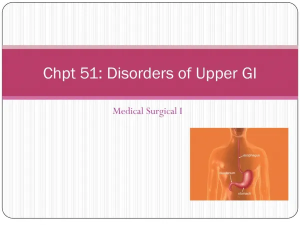
Definitions • Peptic Ulcer Disease: A group of disorders characterized by erosion of the mucosa anywhere in the GI tract that is exposed to the erosive action of acid and pepsin • Erosions (more superficial) may be reepithelialized rapidly by a process called restitution. • Ulcersextend into the deeper layers of the mucosa and require a more complex healing process.
Definitions • Duodenal • Associated with acid output • usually seen in the proximal duodenum [the 1st few cm past pyloric sphincter] • Increased pepsinogen I (parietal cell mass) • Gastric ( in Stomach) • Usually not associated with acid output • occur mainly in the antrum of the stomach
Typesof PUD 1-Non ulcer dyspepsia • Dyspepsia: an imprecise symptom complex that includes: epigastric pain or discomfort, Nausea, belching, bloating. • There is over lap between the symptoms reposted by patients diagnosed with peptic ulcer and dyspepsia
2-NSAID-Induced Ulcers • Symptomatic ulcers occur in only 1% of patients after 3-6 months of NSAIDs • This type of ulcer does not correlate with pain because analgesic action may mask ulcer pain Mechanisms of Injury: • Produce gastric damage by two mechanisms • Direct irritant effect • Systemic effect →inhibition of cyclooxygenase COX-1 → ↓ PG synthesis
PG protect the GI mucosa by maintaining blood flow & stimulating bicarbonate & mucus • Low PG impair the ability of the gastric mucosa to withstand aggressive factors • COX –2 inhibitions is responsible for anti-inflammatory and analgesic effects • Ulcers occur more frequently in the stomach than in the duodenum. • Many are painless • acute, low-dose NSAID-induced lesions usually heal spontaneously
Risk factors • Age > 60 yrs, • High-NSAID dose OR Use of multiple NSAIDs, • Hx of ulcer/complication, • Co-morbid illness, • Concurrent steroid use, • GI sx in past 6 mos requiring discontinuation of the NSAID or the addition of another drug. • Patients taking anticoagulants • High surgical risk or Debilitated patients
3-Adrenocorticosteroids • Association of steroids and PUD is controversial. • Steroids may induce ulcers:↑ gastric acid secretion, ↓ PG production • May delay or inhibit healing of ulcers caused by NSAIDs. • Patients taking steroids considered at high risk • Recent studies: elderly taking both steroids and NSAIDs are at much higher risk of PUD than those receiving either agent alone (related to dose & duration of therapy)
4-Disorders of Acid Hypersecretion • Increased basal & postprandial acid secretion • Mechanisms: • Enhanced sensitivity of the parietal cell to vagal stimulation, • Impaired acid inhibitory mechanisms Zollinger-Ellison Syndrome: caused by a gastrin-secreting tumor of the duodenum or pancreas → marked gastric acid hypersecretion & recurrent peptic ulceration. • 2/3 of tumors are malignant. • Found in ~ 0.1% of patients with duodenal ulcers.
Risk Factors for PUD • Cigarette smoking • Genetic factors • Psychological & physiological stress - multiple trauma, sepsis, neurosurgical problems, ICU stresses • Dietary factors
Chief complaint: Pain (25% are painless) doesn’t always correlate w/presence of acid or ulcer craters Duodenal UlcerGastric Ulcer Most common symptom Most common symptom Sharp, burning, or gnawing Less typical & predictable Occurs 1-3 hrs after meals May occur during a meal Relieved by alkali & food Food offers little/no relief; may precipitate it Usually located in RUQ May extend over a wide area of epigastrum Nocturnal; awakens patient (1-2am)
Clinical presentation of ZE Syndrome • Similar to sever PUD • More persistent • Less responsive to standard therapy • Abdominal pain; most common Sx • Diarrhea: because gastrin inhibits sodium and water reabsorption from intestine
Complications Three Major Complications • Hemorrhage • Perforation • Gastric outlet obstruction • Initially treated conservatively • May require surgery at any time during course of therapy
Hemorrhage • Most common complication of peptic ulcer disease • Develops from erosion of • Granulation tissue found at base of ulcer during healing • Ulcer through a major blood vessel
Perforation • Most lethal complication of peptic ulcer • Commonly seen in large penetrating duodenal ulcers that have not healed and are healed and are • located on posterior mucosal wall • Occurs when ulcer penetrates serosal surface • Size of perforation directly proportional to length of time patient has had ulcer
Gastric Outlet Obstruction • Active ulcer formation is associated with edema, inflammation • Duodenum can predispose to gastric outlet obstruction • ↑ contractile force needed to empty stomach results in hypertrophic stomach wall • Obstruction is not totally due to fibrous scar tissue • Short duration or absence of pain indicative of a malignant obstruction • Vomiting and Constipation are a common complaint
Diagnosis • Visualization of the ulcer defines the disease • 1-Barium Upper GI Study (X-rays) • Can show obstruction, perforation, or penetration • Cannot show bleeding • 30-60% of ulcers can be detected
2-Endoscopy • Most often used • Superior test for diagnosis • More costly & less patient acceptance • Can visualize the ulcer • Can visualize any bleeding • Can treat bleeding • Determines degree of ulcer healing after treatment • Tissue specimens can be obtained to identify and to rule out gastric cancer
Indications for Endoscopy: • New onset dyspepsia in pts > 45 yrs old (to rule out gastric cancer) • Patients with “alarm” symptoms: • Evidence of GI bleeding, anemia, unexplained weight loss, recurrent • vomiting, decreased appetite, easy fullness, dysphagia, abdominal mass, • lymphadenopathy • Patients whose symptoms have failed to respond to initial therapy • Patients with recurrent or difficult to control disease
Laboratory tests • Guaiac test on stool sample • CBC should be done if the patient’s stool is guaiac positive • Acute blood loss: • Red cell indices: RBC, Hct, Hgb, • Normocytic (MCV), Normochromic (MCH) • Chronic blood loss: • Iron deficiency: RBC, Hct, Hgb, MCV, MCH
Tests for GUT Blood • Very important tool for screening in the clinic setting • Hemoccult (stool) • Gastroccult (stomach) • Both are based on the catalytic activity of hemoglobin during the conversion of guiac into a blue pigment in the presence of peroxide
Diagnosis of ZE syndrome • Measurement of gastric acid secretion • >15 mmol/hr in patients with no prior gastric surgery • > 5 mmol/hr in patients with prior gastric surgery • fasting gastrin level > 1000 pg/ml • secretin provocative test: • secretion will cause a marked increase in serum gastrin
H. pylori Testing Indications for H. pylori testing: • New onset dyspepsia in patients < 45 years old (with no alarm symptoms) • Active peptic ulcer disease • PMH of documented peptic ulcer, which has not been tested for H. pylori • Gastric lymphoma
Stool Antigens • A stool antigen enzyme immunoassay has a sensitivity of about 94% and specificity of about 90%. • It should not be used to test for eradication of H. pylori until 6- 8 weeks after therapy.
Gold Standard: Gastric biopsy • The Rapid UreaseTest is the most popular and recommended because of its excellent sensitivity & low cost. • The labeled C breath tests utilize H pylori’s ability to hydrolyze urea into ammonia. • Urea labeled with carbon isotope is administered orally • If organism is present, urea is hydrolyzed & pt will exhale labeled Co2 which can be quantified. • 14C is radioactive – not recommended for children, pregnant women, or for multiple uses in the same person. 13C is non-radioactive
The Serological Test • An excellent initial screen to determine infection because the result is known in 20 min. • H. pylori infection elicits an immune response with an increase in IgG and IgA antibodies. There are commercially available tests that measure IgG antibody. • All of the tests – with the exception of serological testing – may be falsely negative in pts who have taken antibiotics, bismuth compounds, or omeprazole in the recent past.
Therapy Therapeutic Objectives • Relieve pain; control symptoms • Prevent pain recurrence • Prevent further irritation while healing occurs • Remove or treat cause if possible • Promote healing • Prevent complications • Maintain the healed ulcer condition Indices of Therapeutic Effect: A. Pain relief/Absence of pain B. Frequency of antacid use C. Endoscopy (to confirm healing – if symptom response is inadequate
General Treatment Principles: • Establish diagnosis with certainty (poor symptom correlation) • Utilize non-drug modalities, removing potential exacerbating factor Non-drug Therapy: • Avoid alcohol • Stop smoking • Avoid caffeine • Avoid aggravating foods • Reduce stress • Eliminate drug- induced causes (NSAIDs, ASA)
H2-blockers MOA • Selectively and competitively Inhibit the action of histamine on H2 receptors of the parietal cells → reducing basal and stimulated secretion of gastric acid Efficacy Equally Effective for: • Treatment of acute ulcers in the absence of H. pylori • Treatment of NSAID-induced ulcers • Maintenance therapy in patients unable to tolerate a course of antimicrobial therapy to eradicate H. pylori
1-H2-blockers Efficacy • 70-95% heal rate in 4-8 weeks • Standard doses inhibit 50- 80% of 24-hour acid secretion • Equal in efficacy to antacid, and sucralfate therapy • Effective as single therapy in pts with no H pylori infection
Side effects • Safe, Frequency of SE is low • Common SE: • Diarrhea, constipation, Mental confusion, headache, dizziness, drowsiness, and rashes • Confusion/agitation occurs more frequently in elderly, renal & hepatic dysfxn • Similar profile (reported more frequently with cimetidine) • Mental status changes, diarrhea, headache (2-3%) • Rare: bone marrow suppression (thrombocytopenia, agranulocytosis)
Cimetidine: Prolonged use – gynecomastia, erectile dysfxn, sperm ct (anti-androgen) • Ranitidine: Hepatitis • Famotidine: CNS, headache • Nizatidine: Sweating, itching, hepatic
Drug Interaction Profile • Table 27-4 • Cimetidine inhibits oxidative metabolism CYP450 • Magnitude of interaction varies from patient to patient • Generally it will reduce clearance of another drug by 20-30% • Clinically significant in drugs with narrow therapeutic window • Addition of cimetidine may require ↓ dosage of the object drug to avoid increased serum concentration • lidocaine, valium, propranolol, metoprolol, warfarin, phenytoin, theophylline
Other H2-blockers have lower affinity for p450 system; fewer drug interactions: • Cimetidine > ranitidine, nizatidine > famotidine • Cimetidine and ranitidine inhibit renal tubular secretion of procanamide and its metabolite • All 4 drugs can affects absorption and reduce bioavailability of some drugs by requiring gastric PH • Cimetidine ↑ PH →slows dissolution of ketoconazole → ↓ absorption • Cimetidine, ranitidine, nizatidine ↑ absorption of ethanol
Route of Administration and Dosing • See Table 27-1 and Table 1 • Control of night time acid secretion is more effective in healing ulcers • May be given as a single bedtime dose for treatment of DU • For ZES: Not totally effective, require large doses and more frequent dosing • The dose to heal an ulcer – DU=GU • The dose to maintain a healed ulcer –DU=GU • Maintenance dose is cut in half, single daily dose
2-Sucralfate MOA • Protects ulcerated mucosa from aggressive factors • It binds to damaged tissue forming a physical barrier to injury Efficacy • 1 gm 4 times daily as effective as 2 gms twice daily • Requires acid medium to work (don’t combine with H2-blocker) • As effective as H2-blockers in promoting DU healing