Download
1 / 22
230 likes | 600 Views
Screening of Urine Bioassay Samples using a Standard Nuclear Medicine Gamma Camera. Chris Martel Director, RSO Brigham & Women’s Hospital Associate in Radiology Harvard Medical School. Rakesh Kannan, RT(N) Dept. of Nuclear Medicine Brigham & Women’s Hospital. Triage Decisions.

E N D
Screening of Urine Bioassay Samples using a Standard Nuclear Medicine Gamma Camera Chris Martel Director, RSO Brigham & Women’s Hospital Associate in Radiology Harvard Medical School Rakesh Kannan, RT(N) Dept. of Nuclear Medicine Brigham & Women’s Hospital
Research Question • If a radiological incident occurred involving the contamination of large numbers of the public, can the gamma camera be used effectively to screen urine bioassay samples to identify those samples that need to be sent to a lab for further analysis? • What can we measure? / What would we miss?
Materials • Siemens Symbia SPECT/CT • Dual head gamma camera • High energy collimator • Plastic drinking water cups (16 oz.) • Cardboard tray with absorbent pad • Capintec CRC-25R Dose Calibrator • F-18 (FDG) Positron emitter – 511 keV annihilation radiation photons
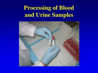
Methods • Cups were filled with about 250 ml of water • Aliquots of F-18 (as measured by dose calibrator) added to cups of water. • Cups placed in cardboard box/absorbent • Box placed on gamma camera head • Standard lung counting protocol selected with F-18 window at 30%. (50% window also available)
Methods • Technologist told to identify the “hot” samples. • Samples and background counted for 3 minutes.
Two hot cups in box others in between Samples were 9-inches apart with a non rad sample between No collimator
Two samples close together (Touching)with no collimator How one draws a region of interest will impact quantitative analysis. Use data to quantify with caution! Suggest using counts per pixel.
Results Efficiency with collimator – 0.01% Efficiency without collimator – 2.2%
IRF-Ingestion IRF-Inhlation
What can we measure? • One can measure 1/10th of an ALI (ingestion or inhalation) for 137Cs beyond 60 days after the event. • For lower energy photon emitters, the attenuation in the sample will be compensated to a degree by the increase in detection efficiency.
What would we miss? • Non (or very low yield) photon emitters • Po-210 • H-3 • C-14 • High energy beta emitters (e.g., Sr/Y-90) may be detectable through brehmstrahlung.
Conclusions • Gamma camera can be used without the collimator to visually screen urine bioassay samples. • Sensitivity appears to be sufficient to make adequate decisions in line with CDGs. • Throughput – 25 samples in total 10 minutes for 150 per hour and 1,200 per 8 hour shift. • Can be increased using scan along bed.
Conclusions • Recommend more robust study on capabilities of gamma camera for screening urine samples. • Different radionuclides • Use camera to scan samples.