Download
1 / 19
190 likes | 326 Views
Integument, Skeletons, movement, Digestion, and Gas Exchange. BIOL240.002 Zoology 3 September 2014. Integument. The skin and its derivative structures Mechanical protection Protection from UV light Protection from infectious organisms Retards moisture loss (terrestrial organisms)

E N D
Integument, Skeletons, movement, Digestion, andGas Exchange BIOL240.002 Zoology 3 September 2014
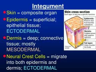
Integument • The skin and its derivative structures • Mechanical protection • Protection from UV light • Protection from infectious organisms • Retards moisture loss (terrestrial organisms) • May be the site of gas exchange
Integument • Single-celled protozoans • Plasma membrane functions as integument • Invertebrates • Epidermis • Single cell layer • Termed hypodermis if it secretes a noncellular outer covering • Ex: Mollusk shell • Ex: Arthropod cuticle • Vertebrates • Stratified epidermis • Stratified dermis underneath • Blood vessels, nerves, connective tissue, fat cells, pigments, etc. Fig. 10.5 p. 190 Fig. 13.2 p. 247 Fig. 20.4 p. 412
Integument • Invertebrate Integumentary Derivatives • Mollusk shells • Arthropod cuticles • Crustaceans: hardened with calcium carbonate • Insects: hardened with the protein sclerotin • Insect wings • Folded over cuticle Fig. 13.50 p. 275
Integument Fig. 16.18 p. 339 • Vertebrates • Dermal bone • Scales • Dermal bone • Epidermal keratin protein • Feathers • Hair • Glands • Claws • Nails • Hooves • Pigments • Chromatophores • Structural color • Horns • Antlers Fig. 18.4 p. 370 Fig. 20.8 p. 414 Fig. 20.4 p. 412
Skeletons • Unicellular protozoans • Cytoskeleton • (Also in all animal cells) • Hydrostatic skeletons • Muscles surround an enclosed fluid • Fluid cannot be compressed • Ex: Nematodes with longitudinal muscles and cuticle • Ex: Annelid worms with circular and longitudinal muscle layers • Also important for certain organs • Ex: Molluscan foot • Ex: Vertebrate tongue Fig. 12.2 p. 234 Fig. 11.2 p. 216 Fig. 10.25 p. 202 Fig. 18.12 p. 375
Skeletons • Exoskeletons • Outer skeleton derived from the integument • Ex: Calcium carbonate cups in which coral polyps sit • Ex: Mollusk shells (clams, snails, etc.) • Ex: Arthropod cuticle Fig. 7.25 p. 146 Fig. 10.1 p. 187 Fig. 13.16 p. 256
Skeletons • Endoskeletons • Internal skeleton, usually derived from connective tissue • Ex: Sponge spicules (calcium carbonate or silicon dioxide) or elastic spongin fibers (protein) • Ex: Squid “pen” • Ex: Echinoderm calcium carbonate ossicles • Ex: Vertebrate cartilage and bone • Bone may be cartilage-replacement bone or dermal bone not preceded by cartilage Fig. 6.9 p. 124 Fig. 10.31 p. 205 Fig. 14.6 p. 296 Fig. 17.10 p. 359
Movement • Protozoan Movement • Pseudopodia—“false feet” • Cytoplasm streams into extended pseudopodia under the action of interacting myosin and actin protein fibers • Cilia • Nine double microtubules around two singles • Triplets at the base • Several cilia beat in coordinated waves • Flagella • Same structure as cilia • One or few, longer, whip-like Fig. 5.5 p. 99 Fig. 5.8 p. 101 Fig. 5.4 p. 98
Movement • Pseudopodia, cilia, and flagella all exist in certain specialized animal cells • Pseudopodia: • Amoeboid sponge archaeocytes • Vertebrate white blood cells • Cilia • Comb jelly rows of cilia • Vertebrate tracheal cells • Flagella • Spermatozoan tails • Sponge feeding cells Fig. 6.8 p. 123 Fig. 7.32 p. 150
Movement • Muscle cells—specialized to contract via protein filaments that slide past one another • Attach to bone, cartilage, shell, cuticle, or surround a fluid-filled cavity • Coelom, digestive tract, blood vessels and heart, etc. • Myosin: thick protein filaments with a head at either end • Actin: thin protein filaments • Six actin filaments surround a myosin filament • Troponin and Tropomyosin: proteins bound to actin which regulate its interaction with myosin • Tropomyosin—blocks myosin head from actin filament • Troponin—holds tropomyosin in place unless bound with Ca++
Animal Nutrition • Raw Materials • Carbohydrates Simple Sugars Carbohydrates • Lipids Glycerol + Fatty Acids Lipids • Proteins Amino Acids Proteins • DNA and RNA Nucleotides DNA and RNA • Energy • Simple Sugars Glycolysis Pyruvate Krebs Cycle • Fattty Acids Acetyl CoA Krebs Cycle • Glycerol Glycolysis Krebs Cycle • Amino Acids Deamination Gylcolysisor Acetyl CoA or Krebs Cycle • Nucleotides Glycolysis Pyruvate Krebs Cycle
Ingestion, Digestion, and Egestion • Protozoans: Pseudopodia, cilia, and flagella are first and foremost feeding structures • Sponge choanocytes: flagella sweep in food • Intracellular digestion • Single-opening gut: mouth for ingestion and egestion • Extracellular digestion of larger prey Fig. 5.9 p. 101 Fig. 6.8 p. 123 Fig. 7.3 p. 133 Fig. 8.8 p. 160
Ingestion, Digestion, and Egestion • Double-opening gut: mouth and anus • Allows division of labor within digestive tract Fig. 7.32 p. 150 Fig. 13.7 p. 251 Fig. 9.9 p. 182
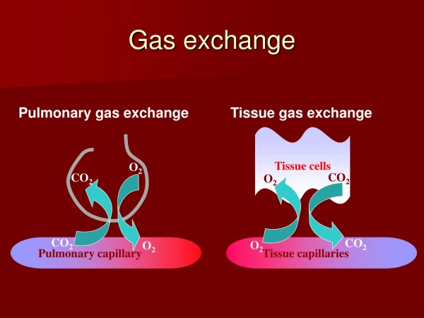
Gas Exchange • Cellular respiration • 1) Glycolysis • 2) Pyruvate Acetyl conversion • 3) Citric acid cycle • 4) Electron transport chain • Last three only happen if O2 is present • CO2 given off during second and third phases • Hence the need for gas exchange • To allow O2 and CO2 exchange via diffusion, repiratory surfaces must be • …moist, to allow diffusion in water rather than air • …permeable, i.e., thin enough to allow passage • …exposed to the water or air • …ventilated, to prevent stagnation of the diffusion gradients • …linked to internal transport
Gas Exchange • Protozoans • Live in water • Permeable plasma membrane is exposed • Ventilation via movement or ciliary currents • Transport to mitochondria via diffusion
Gas Exchange • Very small aquatic animals • Live in water • Epidermal plasma membranes are permeable and exposed • Ventilation via movement or ciliary currents • Transport to other cells via diffusion • Larger aquatic animals • Gills suspended in water • Very fine at point of exchange • Exposed or in exposed chamber • Currents maintained over gills • Linked to circulatory system Fig. 8.19 p. 168 Fig. 10.31 p. 205 Fig. 17.8 p. 358
Gas Exchange • Very small terrestrial animals • Water-filled tracheoles • Open to outside via spiracles • Ventilation via opening/closing of spiracles to direct airflow • Branch inward throughout body • Larger terrestrial animals • Lungs internal to keep moist • Single cell layer at point of exchange • Exposed via tubes (e.g., bronchiiand trachea) • Ventilation via movements (inhalation and exhalation) • Linked to circulatory system Fig. 13.45 p. 272 Fig. 13.7 p. 251 Fig. 19.10 p. 394